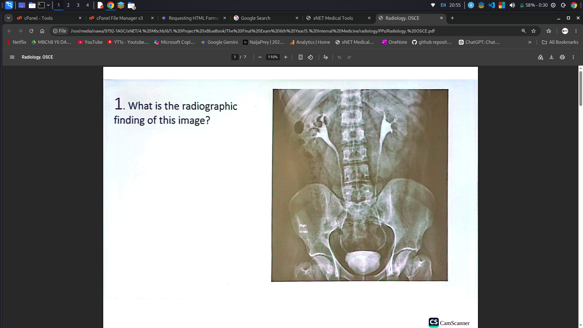
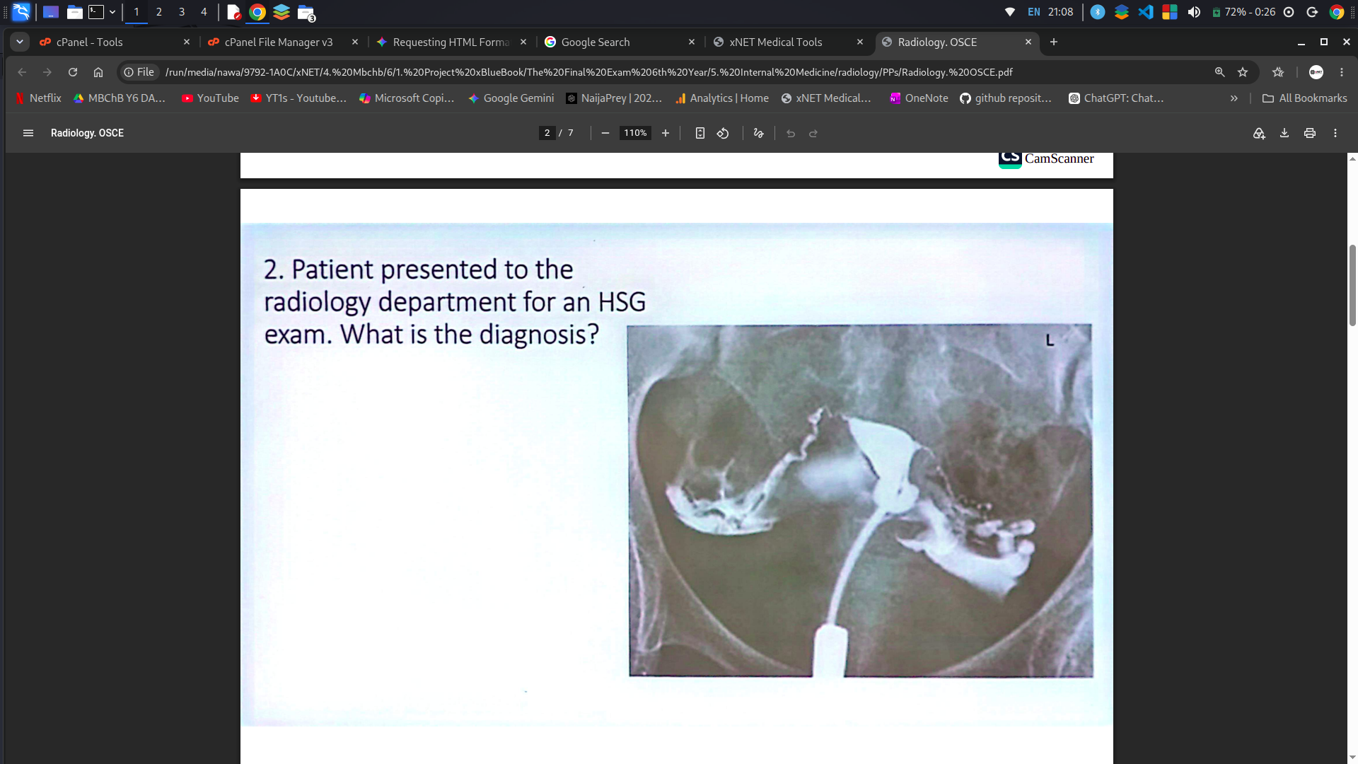
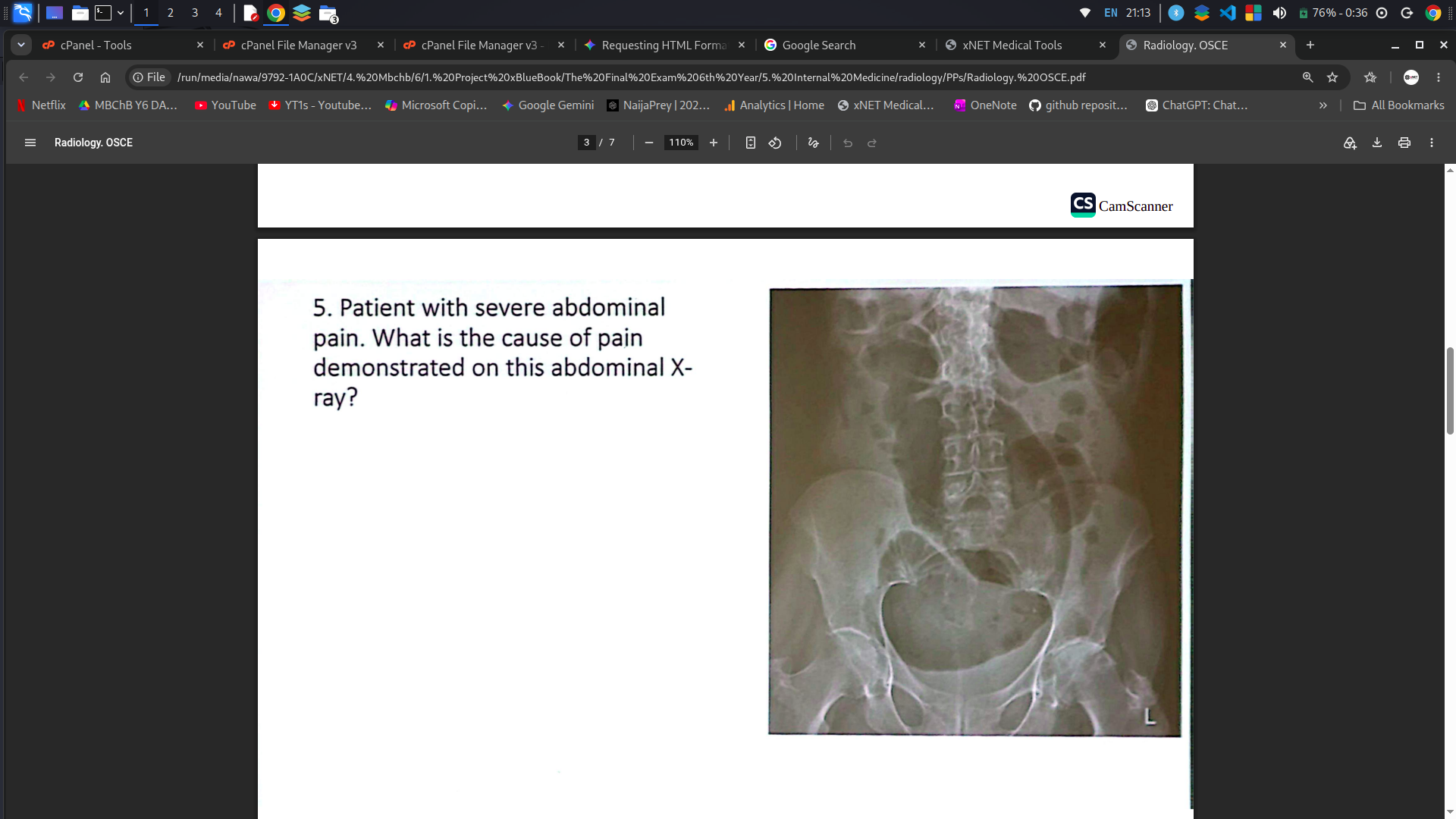
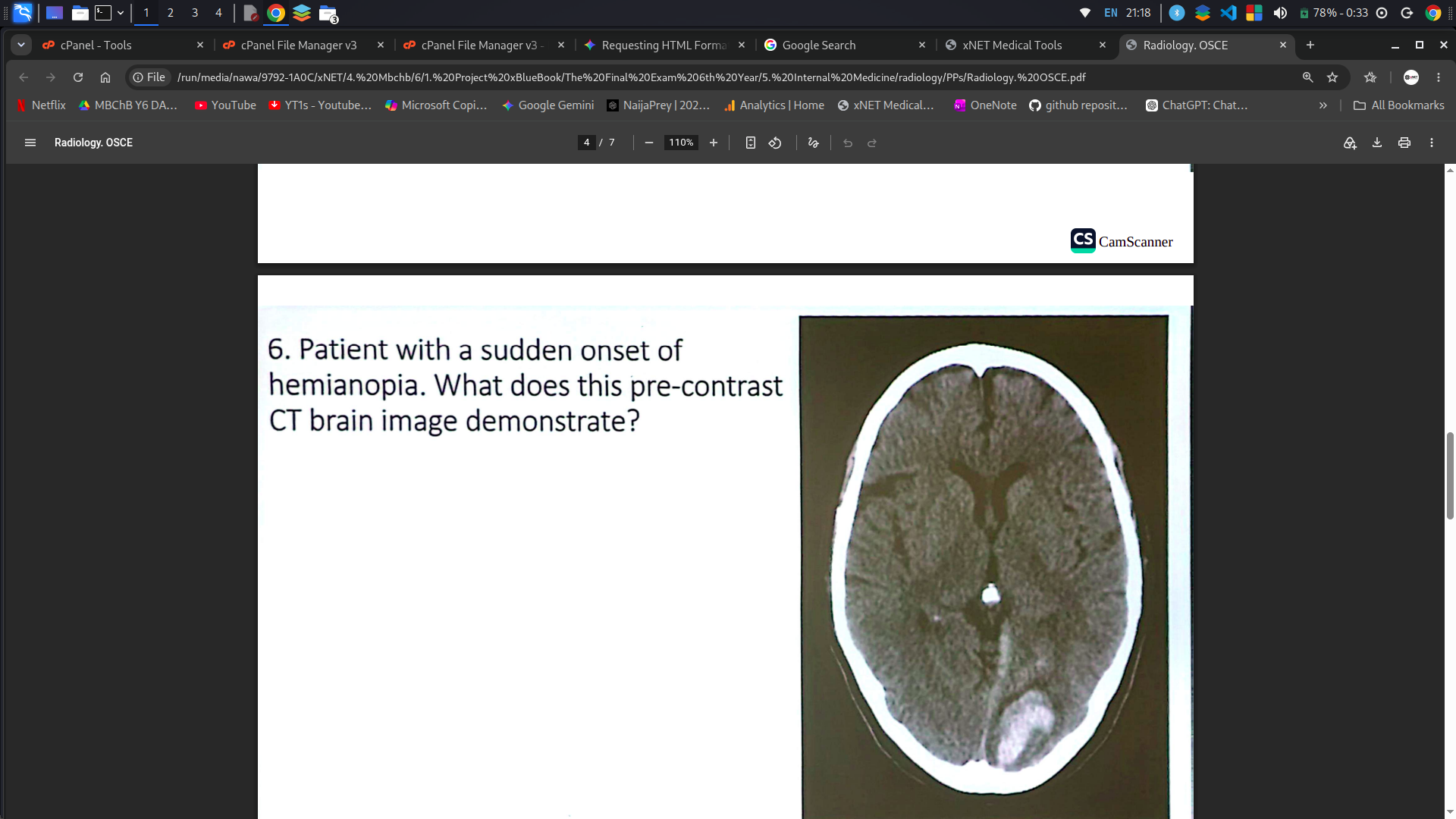
⚠ Disclaimer: Informational use only — not medical advice.
Make a Selection:
Welcome to CT Scan images
CT Scan Findings: Acute Epidural Hematoma
Axial Non-Contrast Head CT Description
An axial non-contrast computed tomography (NCCT) scan of the head demonstrates a classic presentation of an acute epidural hematoma (EDH)[cite: 1].
Morphology: There is a well-circumscribed, high-attenuation (hyperdense) extra-axial collection in the left temporoparietal region displaying a characteristic biconvex or lenticular (lens-shaped) contour[cite: 1].
Dural Borders: The hematoma is confined by skull sutures—where the dura mater firmly attaches to the periosteum—preventing it from crossing suture lines[cite: 1].
Mass Effect: The lesion exerts significant local mass effect on the underlying brain parenchyma, resulting in effacement of adjacent cortical sulci, compression of the ipsilateral lateral ventricle, and subfalcine midline shift toward the right[cite: 1].
Associated Features: An overlying scalp hematoma/soft-tissue swelling is visible along the left calvarium, often associated with an underlying skull fracture that causes laceration of the middle meningeal artery[cite: 1].
Key Diagnostic Summary
Pathophysiology: Typically arises from arterial rupture (most commonly the middle meningeal artery) secondary to temporal bone trauma[cite: 1]. Classic Sign:Biconvex / Lens-shaped hyperdensity bounded by cranial sutures[cite: 1].
Acute Ischemic Stroke
CT Scan Findings: Acute Ischemic Stroke
Axial Non-Contrast Head CT Description
An axial non-contrast head CT scan demonstrates findings characteristic of a large, acute-to-subacute ischemic stroke (cerebral infarction)[cite: 2].
Hypodensity & Vascular Territory: There is a broad, wedge-shaped area of geographic hypoattenuation (darkening) involving the left middle cerebral artery (MCA) territory, affecting both cortical gray matter and underlying subcortical white matter[cite: 2].
Loss of Gray-White Differentiation: Cytotoxic edema causes blurring and loss of the normal contrast between cortical gray matter and white matter architecture across the affected hemisphere[cite: 2].
Mass Effect & Sulcal Effacement: Significant parenchymal swelling results in effacement of the surrounding cortical sulci, partial compression of the ipsilateral (left) lateral ventricle, and subfalcine midline shift toward the contralateral side[cite: 2].
Insular Ribbon & Basal Ganglia Sign: Obscuration of the insular cortex (loss of the insular ribbon) and hypoattenuation within the deep basal ganglia nuclei indicate severe early ischemic injury within the MCA distribution[cite: 2].
Key Diagnostic Summary
Pathophysiology: Acute vascular occlusion (thrombus/embolus) leading to restricted arterial blood flow, cytotoxic edema, and tissue infarction[cite: 2]. Classic Sign:Wedge-shaped cortical/subcortical hypodensity with gray-white boundary loss and local mass effect[cite: 2].
Subarachnoid Hemorrhage
CT Scan Findings: Acute Subarachnoid Hemorrhage
Axial Non-Contrast Head CT Description
Axial non-contrast computed tomography (NCCT) images demonstrate classic features of an acute subarachnoid hemorrhage (SAH)[cite: 3].
High-Attenuation CSF Spaces: There is marked hyperdense (bright) acute blood filling the subarachnoid spaces, normally occupied by dark cerebrospinal fluid (CSF)[cite: 3].
Basal Cistern Involvement: Dense acute hemorrhage diffuses throughout the basal cisterns, creating a characteristic star-shaped hyperdense configuration at the suprasellar cistern[cite: 3].
Sylvian Fissures & Sulci Extension: Extravasated blood outlines the bilateral Sylvian fissures and extends upward into adjacent cortical sulci[cite: 3].
Ventricles & Mass Effect: High-attenuation blood may extend into the third or lateral ventricles, with potential secondary dilatation of the cerebral ventricles indicating early obstructive hydrocephalus[cite: 3].
Key Diagnostic Summary
Pathophysiology: Most commonly non-traumatic, secondary to rupture of an intracranial saccular (berry) aneurysm, often presenting clinically with a sudden "thunderclap" headache[cite: 3]. Classic Sign:Hyperdense acute blood outlining the basal cisterns, Sylvian fissures, and cortical sulci[cite: 3].
Subdural hematoma
CT Scan Findings: Acute Subdural Hematoma
Axial Non-Contrast Head CT Description
An axial non-contrast computed tomography (NCCT) scan of the head reveals typical findings of an acute subdural hematoma (SDH)[cite: 4].
Morphology: There is a prominent hyperdense (bright white) extra-axial collection over the right cerebral convexity showing a characteristic crescentic (concavo-convex) shape[cite: 4].
Anatomical Boundaries: The extra-axial blood tracks freely along the inner surface of the dura, extending across cranial suture lines along the cerebral hemisphere, but restricted by dural reflections (falx cerebri and tentorium cerebelli)[cite: 4].
Mass Effect & Brain Parenchyma Compression: The collection causes significant mass effect, leading to effacement of the overlying cortical sulci, regional compression of the underlying cerebral parenchyma, and compression of the adjacent ipsilateral lateral ventricle[cite: 4].
Associated Features: Soft tissue swelling is noted over the left posterior scalp region[cite: 4].
Key Diagnostic Summary
Pathophysiology: Typically results from shear injury leading to rupture of bridging cortical veins traversing the subdural space, frequently associated with blunt head trauma or rapid deceleration injuries[cite: 4]. Classic Sign:Crescent-shaped hyperdensity crossing suture lines along the cerebral convexity[cite: 4].
Acute Intracerebral Hemorrhage
CT Scan Findings: Acute Intracerebral Hemorrhage
Axial Non-Contrast Head CT Description
An axial non-contrast computed tomography (NCCT) scan of the brain reveals key features of a massive acute intracerebral hemorrhage (ICH) / intraparenchymal hematoma[cite: 5].
High-Attenuation Parenchymal Mass: There is a large, hyperdense (bright white) intra-axial hematoma centered within the deep left basal ganglia and surrounding cerebral parenchyma[cite: 5].
Peri-Hematomal Edema: A peripheral rim of hypoattenuation (darkening) surrounds the high-density blood collection, representing secondary peri-hematomal vasogenic edema and tissue ischemia[cite: 5].
Mass Effect & Structural Displacement: The substantial volume of the acute blood collection causes severe regional mass effect, resulting in complete effacement of the ipsilateral (left) lateral ventricle and a prominent rightward subfalcine midline shift[cite: 5].
Intraventricular Extension: Hyperdense extravasated blood breaks through into the ventricular system, extending into the contralateral ventricular system and third ventricle[cite: 5].
Key Diagnostic Summary
Pathophysiology: Rupture of small penetrating arteries (e.g., lenticulostriate branches of the MCA) most commonly caused by chronic hypertension or cerebral amyloid angiopathy[cite: 5]. Classic Sign:Focal, hyperdense intra-axial collection with surrounding vasogenic edema, ventricular compression, and midline shift[cite: 5].
Cervical spine fracture and dislocation
CT Scan Findings: Cervical Spine Fracture and Dislocation
Sagittal Computed Tomography (CT) Description
A sagittal reformatted CT scan of the cervical spine demonstrates significant trauma-induced mechanical disruption and realignment failure of the mid-cervical vertebrae[cite: 6].
Subluxation / Dislocation: There is anterior displacement (anterolisthesis) of the superior vertebral body relative to the inferior vertebra at the level of injury, indicating disruption of the posterior ligamentous complex and facet joints[cite: 6].
Bony Disruption & Fractures: A fracture line and endplate damage are visible along the affected vertebral body, with potential retropulsion of bone fragments into the spinal canal[cite: 6].
Spinal Canal Compromise: Anterior subluxation combined with fracture retropulsion causes focal narrowing (stenosis) of the central spinal canal, creating high risk for acute spinal cord compression and injury[cite: 6].
Soft Tissue & Alignment Lines: Disruption is evident along the key anatomical alignment lines (anterior longitudinal line, posterior longitudinal line, and spinolaminar line) along with associated prevertebral soft tissue swelling[cite: 6].
Key Diagnostic Summary
Pathophysiology: High-energy hyperflexion, extension, or axial loading trauma (e.g., motor vehicle accidents, falls) resulting in bone fracturing and ligamentous tearing[cite: 6]. Classic Sign:Disruption of smooth sagittal vertebral alignment lines with anterolisthesis, facet dislocation, and canal stenosis[cite: 6].
A coronal contrast-enhanced CT / CT venogram image demonstrates diagnostic features of cerebral venous sinus thrombosis (CVST) involving the superior sagittal sinus[cite: 7].
Empty Delta Sign: A central non-enhancing intraluminal filling defect (representing the intraluminal thrombus) is surrounded by a triangular rim of contrast enhancement along the dura of the superior sagittal sinus[cite: 7].
Venous Sinus Occlusion: Failure of contrast opacification within the thrombosed sinus lumen compared to normal patent venous structures[cite: 7].
Parenchymal Changes: Occlusion of venous drainage can lead to secondary localized edema, venous congestion, or hyperdense hemorrhagic venous infarction[cite: 7].
Unenhanced CT Features (if non-contrast): May demonstrate the dense vein sign or cord sign, appearing as linear high attenuation within the thrombosed venous sinus prior to contrast administration[cite: 7].
Key Diagnostic Summary
Pathophysiology: Thrombotic occlusion of dural venous sinuses or cortical veins leading to impaired venous drainage, elevated intracranial pressure, and potential venous infarction[cite: 7]. Classic Sign:Empty delta sign on contrast CT/CTV and absent flow signal/filling defect within the venous sinus[cite: 7].
Pulmonary embolism
CT Scan Findings: Acute Pulmonary Embolism (PE)
Axial CT Pulmonary Angiogram (CTPA) Description
Axial contrast-enhanced CT pulmonary angiogram (CTPA) images demonstrate pathognomonic features of an acute pulmonary embolism (PE) at the level of the pulmonary artery bifurcation[cite: 8].
Intraluminal Filling Defect: A large, central, non-enhancing intraluminal filling defect (thrombus) is visible in the left pulmonary artery (circled in yellow and zoomed in the close-up inset), surrounding bright intravascular contrast material[cite: 8].
Bilateral Thrombus Burden: Additional smaller filling defects and luminal compromise are present in the right pulmonary artery branches[cite: 8].
Right Ventricular Strain (RV Dilatation): There is noticeable enlargement and dilatation of the right ventricle relative to the left ventricle, indicating acute right ventricular overload/strain[cite: 8].
Interventricular Septal Flattening: The interventricular septum appears flattened and bowed toward the left ventricle secondary to increased right ventricular pressure[cite: 8].
Key Diagnostic Summary
Pathophysiology: Obstruction of the pulmonary arterial tree by a embolized thrombus (most commonly originating from deep vein thrombosis of the lower extremities) leading to increased right ventricular afterload and impaired gas exchange[cite: 8]. Classic Sign:Intraluminal filling defect on CTPA accompanied by right ventricular strain signs (RV enlargement, interventricular septal flattening/bowing)[cite: 8].
Aortic Dissection
CT Scan Findings: Acute Aortic Dissection (Stanford Type A)
Axial Contrast-Enhanced Chest CT Description
An axial contrast-enhanced CT scan of the chest demonstrates pathognomonic features of an acute Stanford Type A aortic dissection involving both the ascending and descending thoracic aorta[cite: 9].
Intimal Flap Visualization: A linear, curvilinear low-attenuation membrane (intimal flap) is clearly visible separating the lumen of the aorta into two distinct vascular channels[cite: 9].
True vs. False Lumen Delineation:
True Lumen: Typically smaller in cross-sectional area, continuous with non-dissected segments, and surrounded by intact intima[cite: 9].
False Lumen: Often larger in caliber due to higher blood volume/pressure over time, containing blood flow within the media layer[cite: 9].
Anatomical Extent: The dissection flap extends throughout the ascending aorta, aortic arch, and into the descending thoracic aorta[cite: 9].
Secondary Signs to Assess: Pericardial effusion or hemopericardium (indicating retrograde propagation/rupture into the pericardial sac), mediastinal hematoma, and involvement of major branch vessels (e.g., brachiocephalic, carotid, or subclavian arteries)[cite: 9].
Key Diagnostic Summary
Pathophysiology: Intimal tear allows high-pressure blood to surge into the aortic media, creating a false lumen that tracks longitudinally along the vessel wall[cite: 9]. Stanford Classification:
Type A: Involves the ascending aorta (surgical emergency)[cite: 9].
Type B: Involves only the descending aorta distal to the left subclavian artery (typically managed medically unless complicated)[cite: 9].
Classic Sign:Curvilinear intimal flap dividing the aortic lumen into true and false channels[cite: 9].
Axial non-contrast (left) and IV contrast-enhanced arterial phase (right) CT images demonstrate critical diagnostic features of a ruptured abdominal aortic aneurysm (AAA)[cite: 10].
Aneurysmal Sac Expansion: A severely dilated infrarenal abdominal aorta measuring approximately 7.5 cm in maximum transverse diameter (aneurysmal threshold $>3\text{ cm}$)[cite: 10].
Aortic Wall Defect & Active Extravasation: A distinct focal disruption/defect is visible along the left posterolateral aortic wall[cite: 10]. The contrast-enhanced arterial phase CT demonstrates a high-attenuation contrast jet (active extravasation) spurting out through this wall defect, confirming ongoing active arterial bleeding[cite: 10].
Retroperitoneal Hematoma: A large, non-uniform acute retroperitoneal hemorrhage/hematoma surrounds the aneurysmal sac and fills the left retroperitoneal space[cite: 10].
Mass Effect & Displacement: The substantial retroperitoneal blood volume causes significant mass effect, resulting in anterior displacement of the left kidney[cite: 10].
Key Diagnostic Summary
Pathophysiology: Progressive weakening and dilatation of the aortic wall (often secondary to atherosclerosis, hypertension, or connective tissue degeneration) leading to transmural wall rupture and life-threatening retroperitoneal hemorrhage[cite: 10]. Classic Sign:Focal aortic wall defect with surrounding retroperitoneal hematoma and active contrast extravasation ("contrast jet")[cite: 10].
Pneumothorax
CT Scan Findings: Acute Right Pneumothorax
Axial Chest CT Description
An axial computed tomography (CT) scan of the chest demonstrates key features of a large right-sided pneumothorax[cite: 11].
Visceral Pleural Line: A sharp, thin, curvilinear radiopaque line (visceral pleura) is clearly visible, separated from the inner aspect of the chest wall[cite: 11].
Pleural Air Space & Absent Markings: There is a large collection of low-attenuation air filling the right pleural cavity[cite: 11]. Complete absence of bronchovascular lung markings is noted peripheral to the visceral pleural line[cite: 11].
Lung Parenchymal Collapse: The underlying right lung parenchyma is partially atelectatic and collapsed inward toward the hilum[cite: 11].
Contralateral Comparison: The non-affected left lung exhibits normal expansion with intact peripheral bronchovascular markings extending to the chest wall[cite: 11].
Key Diagnostic Summary
Pathophysiology: Accumulation of air within the pleural space between the parietal and visceral pleura, disrupting normal negative intrapleural pressure and causing elastic recoil collapse of the underlying lung[cite: 11]. Classic Sign:Visible visceral pleural line with complete absence of peripheral vascular lung markings[cite: 11].
Pneumonia and Consolidation
CT Scan Findings: Pneumonia & Lobar Consolidation
Axial Chest CT Description
An axial computed tomography (CT) scan of the chest in lung window settings demonstrates definitive features of acute pneumonia with lobar consolidation involving the left lung[cite: 12].
Dense Parenchymal Consolidation: There is a confluent region of marked high attenuation (opacification) completely filling the lower lobe and peripheral regions of the left lung[cite: 12].
Air Bronchograms: Branching, low-attenuation air-filled conductive bronchi are clearly visualized coursing through the dense, fluid-filled consolidated lung tissue (circled region)[cite: 12].
Ground-Glass Attenuation: Peripheral areas adjacent to dense consolidation exhibit hazy, increased attenuation with preserved underlying vascular structures, indicative of partial alveolar filling and inflammatory exudate[cite: 12].
Contralateral Comparison: The right lung parenchyma appears normal and fully aerated with clear bronchovascular branching[cite: 12].
Key Diagnostic Summary
Pathophysiology: Alveolar spaces fill with inflammatory exudate, pus, and cellular debris secondary to infectious agents (e.g., Streptococcus pneumoniae), replacing normal air spaces while leaving large conducting airways patent[cite: 12]. Classic Sign:Dense parenchymal opacification containing branching air bronchograms[cite: 12].
Cardiomegaly
CT Scan Findings: Cardiomegaly and Pericardial Effusion
Axial Chest CT Description
An axial contrast-enhanced CT scan of the chest demonstrates key structural abnormalities involving the heart and surrounding pericardial sac[cite: 13].
Cardiac Enlargement (Cardiomegaly): Marked enlargement of the cardiac silhouette relative to the transverse thoracic diameter, with diffuse dilatation of the ventricular and atrial chambers[cite: 13].
Pericardial Effusion: A fluid-density collection surrounds the heart within the pericardial sac (indicated by the white arrow on the right anterior pericardial boundary)[cite: 13].
Surrounding Parenchymal Findings: Associated bibasilar dependent fluid collections/atelectasis and left-sided pleural changes secondary to mass effect from the enlarged cardiac silhouette[cite: 13].
Key Diagnostic Summary
Pathophysiology: Compensatory ventricular remodeling, myocardial hypertrophy, or chamber dilatation secondary to heart failure, cardiomyopathy, valvular disease, or systemic hypertension, frequently accompanied by pericardial fluid accumulation[cite: 13]. Classic Sign:Enlarged transverse cardiothoracic dimension with surrounding fluid-density circumferential pericardial space[cite: 13].
Acute Appendicitis
CT Scan Findings: Acute Appendicitis
Axial Abdominal CT Description
An axial contrast-enhanced CT scan of the abdomen and pelvis demonstrates key diagnostic features of acute appendicitis in the right lower quadrant[cite: 14].
Appendiceal Dilatation: The tubular appendix is abnormally enlarged, measuring 8.2 mm in outer diameter (abnormal threshold $>6\text{ mm}$)[cite: 14].
Wall Thickening & Hyperenhancement: The appendiceal wall shows prominent circumferential thickening and bright post-contrast hyperenhancement secondary to acute mucosal inflammation[cite: 14].
Periappendiceal Fat Stranding: Increased attenuation and hazy linear density (fat stranding) surround the inflamed appendix in the right iliac fossa, indicating localized inflammatory exudate[cite: 14].
Appendicolith: A focal hyperdense, calcified density (appendicolith / fecalith) is clearly identified at the base/lumen of the appendix[cite: 14].
Key Diagnostic Summary
Pathophysiology: Luminal obstruction (commonly by an appendicolith) leading to mucosal secretion, elevated intraluminal pressure, venous congestion, ischemia, and bacterial overgrowth[cite: 14]. Classic Sign:Dilated tubular structure ($>6\text{ mm}$) in the RLQ with wall hyperenhancement, periappendiceal fat stranding, and potential appendicolith[cite: 14].
Acute Diverticulitis
CT Scan Findings: Acute Diverticulitis
Axial & Coronal Abdominal CT Description
Axial and coronal contrast-enhanced CT images of the abdomen and pelvis demonstrate key diagnostic features of acute diverticulitis centered in the sigmoid colon[cite: 15].
Focal Colonic Wall Thickening: There is prominent, asymmetric segmental wall thickening of the sigmoid colon[cite: 15].
Colonic Outpouchings (Diverticula): Multiple air- or fluid-filled outpouchings (diverticula) are visible arising from the outer contour of the colonic wall[cite: 15].
Pericolic Fat Stranding: Extensive hazy, increased attenuation (fat stranding) is present in the surrounding pericolic fat, representing acute inflammatory changes and edema[cite: 15].
Anatomical Distribution: Findings are predominantly located in the lower left quadrant involving the sigmoid colon, which is the most common site for diverticular disease[cite: 15].
Key Diagnostic Summary
Pathophysiology: Micro- or macro-perforation of a colonic diverticulum (outpouching of mucosa and submucosa through muscular layer defects), leading to localized pericolic inflammation and infection[cite: 15]. Classic Sign:Segmental colonic wall thickening and multiple diverticula with surrounding pericolic fat stranding, most commonly in the sigmoid colon[cite: 15].
Bowel Obstruction
CT Scan Findings: Bowel Obstruction (SBO/LBO)
Axial & Coronal Abdominal CT Description
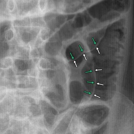
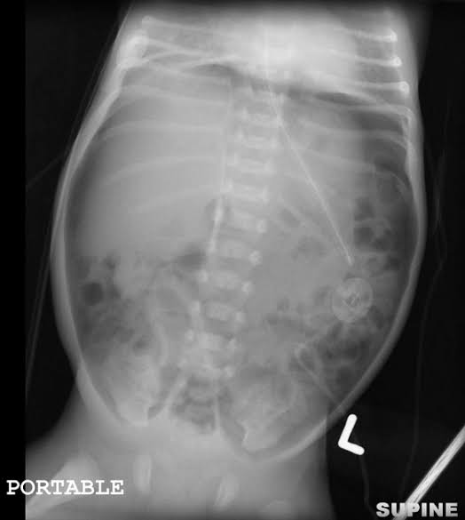
Axial and coronal CT images of the abdomen and pelvis demonstrate definitive features of a mechanical bowel obstruction[cite: 16].
Proximal Bowel Dilatation: Multiple fluid- and gas-distended small/large bowel loops are visible proximal to the site of obstruction[cite: 16].
Air-Fluid Levels: Prominent horizontal air-fluid levels are clearly demonstrated within the dilated proximal bowel loops on both axial and coronal projections[cite: 16].
Transition Point: A distinct point of caliber change (transition point) is identified, where dilated proximal bowel abruptly transforms into decompressed distal bowel[cite: 16].
Distal Bowel Collapse: The distal bowel loops beyond the transition point are collapsed/decompressed due to the lack of downstream passage of luminal contents[cite: 16].
Key Diagnostic Summary
Pathophysiology: Mechanical blockage of the intestinal lumen prevents normal forward passage of gas, fluid, and alimentary contents, leading to proximal distension, intraluminal fluid accumulation, and distal collapse[cite: 16]. Etiology by Classification:
Small Bowel Obstruction (SBO): Most commonly caused by post-operative adhesions, incarcerated hernias, or neoplasms[cite: 16].
Large Bowel Obstruction (LBO): Most commonly caused by colorectal carcinoma, volvulus, or severe diverticulitis[cite: 16].
Classic Sign:Dilated proximal bowel loops with air-fluid levels terminating at a clear transition point leading to collapsed distal bowel[cite: 16].
Axial and coronal CT images of the abdomen and pelvis demonstrate characteristic radiologic signs of a pneumoperitoneum secondary to a perforated viscus[cite: 17].
Extraluminal Free Air in Non-Dependent Areas: Low-attenuation free gas is identified rising to non-dependent peritoneal spaces, specifically along the anterior abdominal wall and within the subdiaphragmatic spaces[cite: 17].
Subdiaphragmatic Crescentic Collections: Prominent crescent-shaped pockets of gas are clearly visualized under the diaphragm (notably seen on both axial and coronal projections)[cite: 17].
Free Air Outlining Mesentery & Bowel Loops: Pockets and streaks of extraluminal air track between mesentery and outline outer bowel walls[cite: 17].
Search for Etiology / Source: Critical CT evaluation involves inspecting for localized fluid collections, bowel wall thickening, or focal defects indicative of the primary site of perforation (e.g., peptic ulcer, perforated appendicitis, diverticulitis, or trauma)[cite: 17].
Key Diagnostic Summary
Pathophysiology: Loss of integrity/transmural perforation of a hollow gastrointestinal organ allows luminal gas and contents to escape into the peritoneal cavity[cite: 17]. Common Causes: Perforated peptic ulcer disease, complicated diverticulitis, gangrenous appendicitis, ischemic bowel, or penetrating/blunt abdominal trauma[cite: 17]. Classic Sign:Free extraluminal gas accumulating in non-dependent areas, particularly crescentic subdiaphragmatic or anterior abdominal wall collections[cite: 17].
Bowel ischemia
Detailed Information for Annotated CT Image
This image is a contrast-enhanced computed tomography (CT) scan of an abdomen with multiple anatomical and pathological findings annotated. A scale in centimeters is visible on the right side.
Ischemic Bowel Loop (Hypoenhancing): A pointer on the left side of the image highlights this finding within the liver region, suggesting reduced blood flow to a bowel loop. (Coordinates: 104, 219)
Hyperemic Bowel Loop (Hyperenhancing): A pointer on the right side highlights an area in the bowel that appears more brightly enhanced, indicating increased blood flow, which may be a compensatory response or part of an inflammatory process. (Coordinates: 706, 95)
Pneumatosis Intestinalis: Annotated in the same vicinity as the hyperemic bowel loop on the right, this term describes the presence of gas within the bowel wall, often a sign of bowel wall injury or ischemia. (Coordinates: 889, 237)
Portal Venous Gas (in Liver): A pointer near the center highlights gas within the portal venous system in the liver, a finding often associated with significant bowel ischemia and potentially serious conditions. (Coordinates: 900, 396)
Ischemic Bowel Loop (Intestinalis): Located in the lower-left section, this annotation likely refers to ischemic bowel loops in the intestinal region, further suggesting widespread bowel ischemia. (Coordinates: 85, 715)
Mesenteric Artery: Identified near the center-right, this structure is a major blood vessel supplying blood to the intestines. (Coordinates: 893, 622)
Mesenteric Vein: Also in the center-right area, this vein drains blood from the intestines. (Coordinates: 888, 792)
Target sign
Detailed Information for Intussusception CT Scan
This image is an axial view of a contrast-enhanced computed tomography (CT) scan of the mid-abdomen demonstrating a classic "target sign" characteristic of intussusception[cite: 2].
Annotated Findings & Features
Target Sign (Concentric Rings): A distinct concentric ring pattern in the mid-jejunal region representing telescoping bowel layers[cite: 2].
Intussusceptum: The innermost part of the invagination containing the proximal bowel segment along with its mesentery and vessels[cite: 2].
Intussuscipiens: The outermost receiving distal bowel loop that encompasses the invaginated segment[cite: 2].
Mesenteric Fat and Vessels within Intussusception: Mesenteric structures invaginated between the intussusceptum and intussuscipiens[cite: 2].
Normal Appearing Bowel: Unaffected, normal-appearing bowel loops are visible adjacent to the obstruction[cite: 2].
Summary of Findings
Axial view of the mid-abdomen reveals a distinct target sign in the mid-jejunal region, consistent with an intussusception, where a proximal segment (intussusceptum) is invaginated into a distal segment (intussuscipiens)[cite: 2]. Multiple other bowel loops are dilated, suggesting partial obstruction[cite: 2].
Detailed Information for Acute Cholecystitis CT Scan
This image is an axial computed tomography (CT) scan of the upper abdomen depicting classic imaging features of acute cholecystitis[cite: 3].
Annotated Imaging Findings
Distended Gallbladder: Marked enlargement of the gallbladder lumen[cite: 3].
Thickened Wall (>3 mm): Measured thickening of the gallbladder wall exceeding 3 mm, characteristic of gallbladder inflammation[cite: 3].
Radiopaque Gallstones and Sludge: Hyperdense calcified stones and dense fluid/sludge visualized within the gallbladder neck/lumen[cite: 3].
Pericholecystic Fat Stranding: Increased density and inflammatory haziness in the fat surrounding the gallbladder wall[cite: 3].
Pericholecystic Fluid Collection: Fluid accumulation adjacent to the gallbladder wall secondary to acute inflammation[cite: 3].
Anatomical Landmarks: Prominently labeled surrounding structures include the Liver and the Portal Vein[cite: 3].
h1>Detailed Information for Acute Pancreatitis CT Scan
This image is an axial contrast-enhanced computed tomography (CT) scan of the upper abdomen demonstrating typical features of acute necrotizing pancreatitis and peripancreatic involvement[cite: 4].
Annotated Pathological & Imaging Findings
Necrotic Pancreatic Parenchyma: Non-enhancing areas of the pancreatic tissue indicating parenchymal necrosis[cite: 4].
Peripancreatic Fluid Collection: Fluid collections surrounding the pancreas, visible in multiple areas including near the pancreatic tail and anterior/lateral regions[cite: 4].
Peripancreatic Fat Stranding: Inflammatory haziness and linear attenuation within the retroperitoneal fat around the pancreas[cite: 4].
Annotated Anatomical Landmarks
Pancreas: The primary organ involved, centered in the upper retroperitoneum[cite: 4].
Liver: Labeled prominently in the upper right side of the image (anatomical right lobe and left lobe)[cite: 4].
Stomach: Contrast-filled structure anterior to the body and tail of the pancreas[cite: 4].
Duodenum: Visualized adjacent to the head of the pancreas[cite: 4].
Aorta: The major arterial vessel centered anterior to the spine[cite: 4].
L. Kidney: The left kidney located posterior to the pancreatic tail[cite: 4].
Spleen: Visible in the upper left abdomen adjacent to the tail of the pancreas[cite: 4].
Hepatic steatosis
Detailed Information for Hepatic Steatosis CT Scan
This image displays a non-contrast CT scan evaluating Hepatic Steatosis (Fatty Liver), featuring side-by-side axial views with attenuation measurements and diagnostic criteria[cite: 5].
Annotated Quantitative Measurements
LIVER ROI: Region of interest measured in the liver parenchyma showing an average attenuation of 35 HU[cite: 5].
SPLEEN ROI: Region of interest measured in the spleen showing an average attenuation of 52 HU[cite: 5].
Detailed Inset Features & Annotations
Liver Parenchyma: Diffuse hypoattenuation: The liver tissue exhibits diffusely decreased attenuation relative to normal parenchyma and the spleen[cite: 5].
Spleen: Reference organ: Used as an internal control for attenuation comparison on non-contrast imaging[cite: 5].
Attenuation Difference Metrics: Highlighted text indicates values comparing liver to spleen attenuation (>10 HU and ~10 HU criteria)[cite: 5].
Diagnostic Conclusion
Diagnosis: Moderate to Severe Hepatic Steatosis[cite: 5].
CT Criteria Met: Hepatic attenuation is more than 10 HU lower than the splenic attenuation on non-contrast imaging[cite: 5].
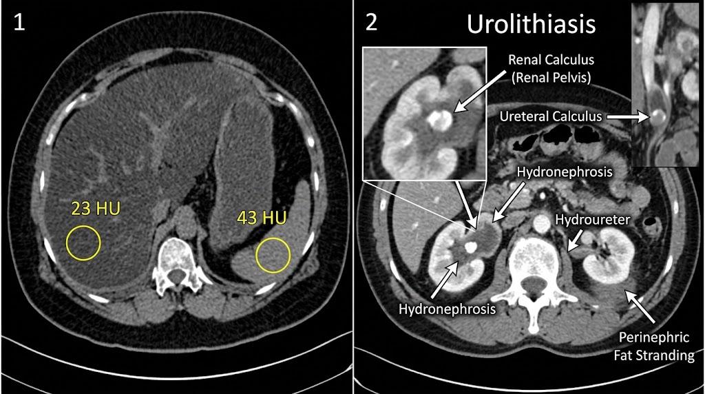
Renal calculi
Detailed Information for Urolithiasis CT Scan
This image presents a two-panel comparative CT assessment detailing non-contrast liver/spleen attenuation measurements (Panel 1) alongside comprehensive imaging findings of urolithiasis and urinary tract obstruction (Panel 2)[cite: 6].
Panel 1: Non-Contrast Attenuation Assessment
Liver Attenuation (23 HU): Region of interest in the liver measuring 23 Hounsfield Units, demonstrating significant hepatic hypoattenuation consistent with hepatic steatosis[cite: 6].
Spleen Attenuation (43 HU): Region of interest in the spleen measuring 43 Hounsfield Units serving as the reference organ baseline[cite: 6].
Panel 2: Urolithiasis & Obstructive Features
Renal Calculus (Renal Pelvis): Hyperdense calculus located within the renal pelvis, highlighted in the magnified inset[cite: 6].
Ureteric Calculus: Obstructing hyperdense stone demonstrated within the ureter in the coronal insert view[cite: 6].
Hydronephrosis: Marked dilatation of the renal pelvicalyceal system visible in both kidneys[cite: 6].
Hydroureter: Dilatation of the left ureter secondary to downstream urinary obstruction[cite: 6].
Perinephric Fat Stranding: Linear haziness within the perinephric fat surrounding the left kidney, indicating acute inflammation or edema from urinary tract obstruction[cite: 6].
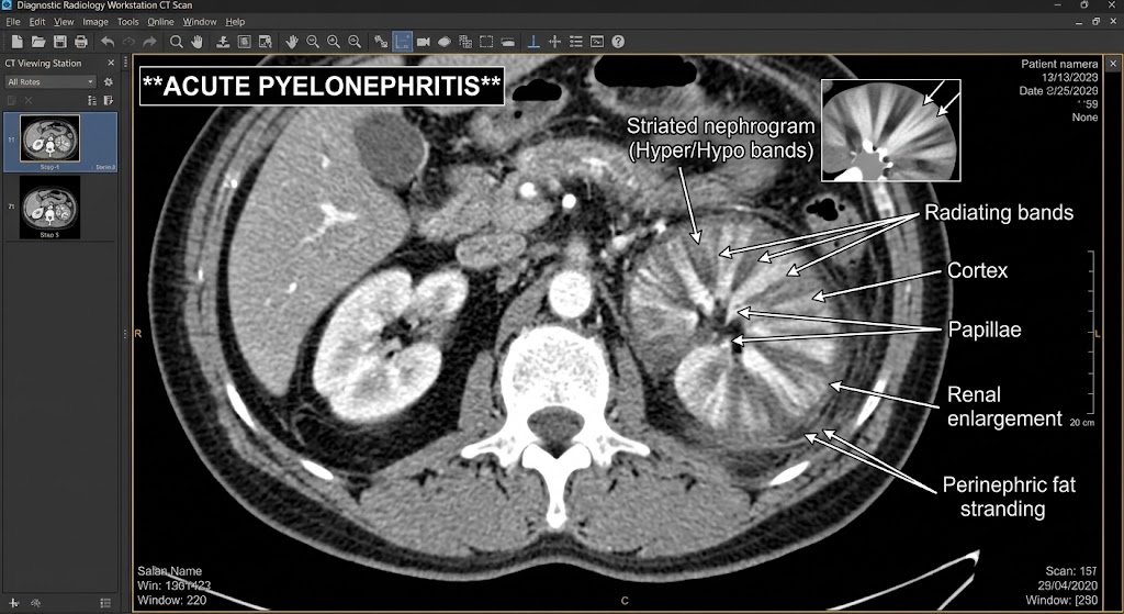
Acute pyelonephritis
Detailed Information for Acute Pyelonephritis CT Scan
This image is a contrast-enhanced axial computed tomography (CT) scan of the abdomen demonstrating classic features of acute pyelonephritis involving the left kidney[cite: 7].
Annotated Imaging Findings
Striated Nephrogram (Hyper/Hypo bands): Characteristic alternating hyperdense and hypodense linear bands extending across the renal parenchyma, highlighted in both the primary CT view and top-right inset[cite: 7].
Radiating Bands: Distinct wedge-shaped or linear bands radiating outward from the medulla towards the cortex, reflecting tubular obstruction and interstitial edema[cite: 7].
Renal Enlargement: Significant swelling and enlargement of the affected left kidney compared to the right kidney[cite: 7].
Perinephric Fat Stranding: Inflammatory linear attenuation and hazy density surrounding the posterior aspect of the left kidney[cite: 7].
Annotated Anatomical Features
Cortex: Outer region of the kidney showing alternating attenuation bands[cite: 7].
Papillae: Innermost projections of the renal pyramids targeted near the center of the radiating bands[cite: 7].
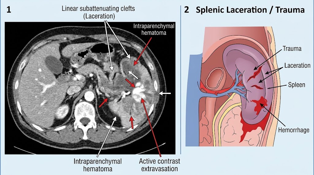
Detailed Information for Splenic Laceration / Trauma
This image features a two-panel presentation consisting of a contrast-enhanced axial CT scan (Panel 1) and a corresponding anatomical illustration (Panel 2) depicting splenic trauma, lacerations, hematoma formation, and active vascular contrast extravasation[cite: 8].
Panel 1: Axial CT Imaging Findings
Linear Subattenuating Clefts (Laceration): Hypodense linear defects extending through the splenic parenchyma representing traumatic lacerations[cite: 8].
Intraparenchymal Hematoma: Non-enhancing low-attenuation regions within the splenic tissue corresponding to localized parenchymal hematomas[cite: 8].
Active Contrast Extravasation: Bright, high-attenuation focus within the spleen indicating active vascular arterial bleeding[cite: 8].
Panel 2: Anatomical Diagram Findings
Trauma: Illustrates the mechanism of injury to the upper left quadrant[cite: 8].
Laceration: Visualized as irregular tears across the splenic capsule and parenchyma[cite: 8].
Spleen: Depicts the affected organ and its vascular supply[cite: 8].
Hemorrhage: Shows active internal bleeding and subcapsular/perisplenic blood collection[cite: 8].
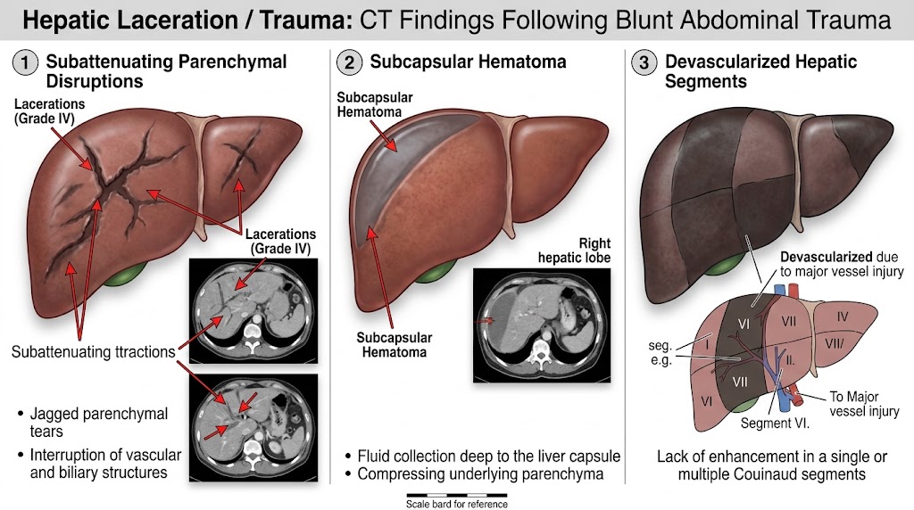
Hepatic laceration
Detailed Information for Hepatic Laceration / Trauma: CT Findings Following Blunt Abdominal Trauma
This image illustrates three key patterns of hepatic trauma CT findings following blunt abdominal trauma, featuring anatomical diagrams paired with corresponding CT scan images and bulleted descriptions[cite: 9].
1. Subattenuating Parenchymal Disruptions
Lacerations (Grade IV): Deep branching hypodense defects across the liver parenchyma[cite: 9].
Jagged parenchymal tears: Irregular linear hypodensities traversing liver tissue[cite: 9].
Interruption of vascular and biliary structures: Traumatic injury to intrahepatic vessels and bile ducts[cite: 9].
2. Subcapsular Hematoma
Subcapsular Hematoma: Crescentic fluid accumulation situated deep to the liver capsule and peripheral to the parenchyma, causing flattening or compression of underlying liver tissue[cite: 9].
Right Hepatic Lobe Involvement: Demonstrated along the anterolateral margin of the right liver lobe on CT[cite: 9].
Fluid collection deep to the liver capsule: Characterized on non-contrast or early post-contrast CT[cite: 9].
Compressing underlying parenchyma: Mass effect on adjacent liver tissue[cite: 9].
3. Devascularized Hepatic Segments
Devascularized due to major vessel injury: Non-enhancing, geographic areas corresponding to specific Couinaud liver segments (e.g., Segment VI) secondary to main or branch vessel transection/thrombosis[cite: 9].
To Major Vessel Injury: Schema illustrating injury to segmental hepatic vessels[cite: 9].
Lack of enhancement in a single or multiple Couinaud segments: Key radiological sign of parenchymal devascularization[cite: 9].
Pelvic fracture
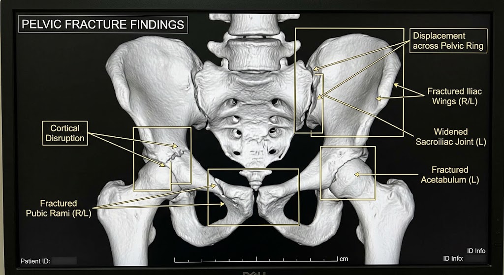
Detailed Information for Pelvic Fracture Findings
This image presents a 3D-reconstructed CT rendering of the bony pelvis demonstrating severe complex pelvic trauma with multiple fractures, joint diastasis, and ring disruption[cite: 10].
Annotated Pathological Findings
Cortical Disruption: Break in the outer bone cortex along the right iliac/acetabular region[cite: 10].
Fractured Pubic Rami (R/L): Disrupted fracture lines involving the superior and inferior pubic rami[cite: 10].
Displacement across Pelvic Ring: Significant displacement and malalignment extending across the posterior pelvic ring structures[cite: 10].
Fractured Iliac Wings (R/L): Fracture lines extending through the iliac wing[cite: 10].
Widened Sacroiliac Joint (L): Diastasis and widening of the left sacroiliac joint space[cite: 10].
Fractured Acetabulum (L): Articular fracture involvement of the left acetabular cavity[cite: 10].
Necrotizing fasciitis
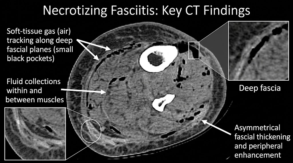
Detailed Information for Necrotizing Fasciitis CT Scan
This image presents a cross-sectional CT scan illustrating key radiological features of necrotizing fasciitis within the limb soft tissues, complete with high-magnification inset views[cite: 11].
Annotated CT Findings
Soft-tissue gas (air) tracking along deep fascial planes (small black pockets): Multiple low-attenuation air pockets dissected along deep fascial borders[cite: 11].
Fluid collections within and between muscles: Hypodense fluid accumulations extending through intermuscular and intramuscular compartments[cite: 11].
Deep fascia: Highlighted in the top-right magnified inset showing gas tracking along the deep fascial layer[cite: 11].
Asymmetrical fascial thickening and peripheral enhancement: Marked thickening of the fascial layers with surrounding inflammatory changes, as well as an inset demonstrating localized soft-tissue alterations[cite: 11].
Abscess formation
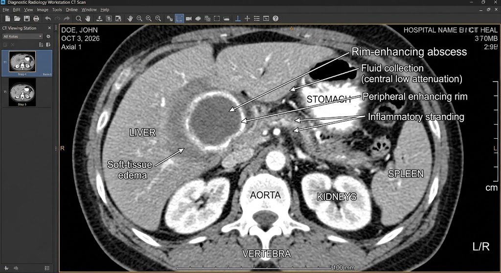
Detailed Information for Rim-Enhancing Abscess CT Scan
This image is a contrast-enhanced axial computed tomography (CT) scan of the upper abdomen demonstrating a well-defined hepatic abscess with classic imaging features[cite: 12].
VERTEBRA: Central osseous landmark along the posterior margin[cite: 12].
DVT
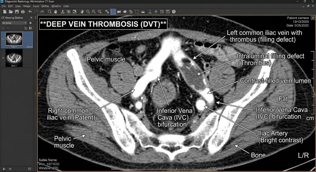
Detailed Information for Deep Vein Thrombosis (DVT) CT Scan
This image is a contrast-enhanced axial computed tomography (CT) scan of the pelvis demonstrating key features of Deep Vein Thrombosis (DVT) within the pelvic venous vasculature[cite: 13].
Annotated Pathological Findings
Left common iliac vein with thrombus (filling defect): Marked expansion and opacification defect within the left common iliac vein[cite: 13].
Welcome to community medicine PastPapers for 6th year Mbchb
Make a Selection:
Question Text:
1. A study conducted at UTH examines anxiety levels of patients before and after receiving a new therapy. On the first trial, a sample of 25 patients shows a mean anxiety score of 30 before therapy (SD=5) and a mean score of 25 after therapy (SD=4). In the second trial, they got a population mean of 200 and the average of 185 with a standard deviation of 15 on a sample of 40 patients. Can you test the hypothesis of this study? (10 MARKS)[cite: 1]
Answer & Solution:
Part 1: Trial 1 (Paired / Two-Sample Comparison)
For Trial 1 ($n = 25$), anxiety levels are evaluated pre- and post-therapy[cite: 1].
Null Hypothesis ($H_0$): $\mu_{\text{before}} - \mu_{\text{after}} = 0$ (There is no difference in mean anxiety score before and after therapy)[cite: 1].
Alternative Hypothesis ($H_1$): $\mu_{\text{before}} - \mu_{\text{after}} \neq 0$ (There is a significant difference in anxiety score after therapy)[cite: 1].
Mean Difference ($\Delta \bar{x}$): $30 - 25 = 5$[cite: 1].
Part 2: Trial 2 (One-Sample Hypothesis Test)
For Trial 2, sample mean $\bar{x} = 185$, hypothesized population mean $\mu_0 = 200$, sample standard deviation $s = 15$, and sample size $n = 40$[cite: 1].
Null Hypothesis ($H_0$): $\mu = 200$[cite: 1]
Alternative Hypothesis ($H_1$): $\mu \neq 200$[cite: 1]
Conclusion: Since $|t| = 6.32$ exceeds the critical value (e.g., $t_{\text{critical}} \approx 2.023$ at $\alpha = 0.05, df = 39$), we reject the null hypothesis ($H_0$). The therapy resulted in a statistically significant reduction in anxiety scores[cite: 1].
1. What are the key differences between applied research and basic research, and how do these differences impact their respective goals and methodologies? (10 Marks)[cite: 2]
Answer:
Key Differences, Goals, and Methodologies:
Primary Purpose / Goal:
Basic Research (Pure / Fundamental): Aimed at expanding fundamental knowledge, understanding underlying principles, and discovering basic laws of nature without immediate practical application.
Applied Research: Directed toward solving specific, practical, real-world problems or developing practical applications, intervention strategies, and practical technologies.
Methodological Differences:
Basic Research: Conducted primarily in highly controlled environments (such as laboratories or theoretical simulations) to establish causality, universal generalizability, and theoretical framework consistency.
Applied Research: Conducted in real-world environments or field settings (e.g., clinical trials, community interventions, organizational setups) to address contextual variables and yield immediate actionability.
Time Horizon & Outcome: Basic research yields theoretical constructs and baseline scientific literature that may take decades to commercialize, whereas applied research produces actionable solutions, policies, treatments, or patents meant for immediate implementation.
Question 2:
2. Can you provide examples of how applied research has successfully addressed real-world problems in industries such as healthcare? (10 Marks)[cite: 2]
Answer:
Examples of Applied Research in Healthcare:
Vaccine and Therapeutic Development: Translating basic microbiological research into clinical trials to develop target-specific vaccines (e.g., mRNA vaccine platforms for SARS-CoV-2, malaria vaccines like RTS,S/AS01 and R21/Matrix-M), directly reducing morbidity and mortality in global populations.
Implementation of Diagnostic Protocols: Applying rapid diagnostic tests (RDTs) for malaria, HIV, and tuberculosis at point-of-care primary health facilities, drastically reducing diagnosis turnaround times and ensuring rapid therapeutic initiation in resource-limited settings.
Quality Improvement & Infection Control: Investigating post-operative infection rates in surgical wards to implement standardized WHO surgical safety checklists and hygiene protocols, resulting in lower hospital-acquired infection (HAI) rates.
Epidemiological Disease Control: Utilizing applied epidemiological studies to map vector distribution (e.g., Anopheles mosquitoes) and evaluating the real-world effectiveness of Insecticide-Treated Nets (ITNs) or Indoor Residual Spraying (IRS) in reducing endemic disease transmission.
Question 3:
3. How do ethical considerations play a role in the design and implementation of applied research studies, especially when dealing with sensitive or potentially harmful subjects? (10 Marks)[cite: 2]
Answer:
Role of Ethical Considerations in Applied Research:
Informed Consent and Voluntariness: Ensuring participants receive clear, comprehensible, and culturally appropriate information regarding the study's purpose, risks, and benefits, while ensuring participation remains entirely voluntary without coercion.
Beneficence and Non-Maleficence: Maximizing potential benefits while minimizing physical, psychological, social, or legal harm. Designing protocols with safety stop-rules and risk mitigation measures for high-risk interventions.
Protection of Vulnerable Populations: Applying extra safeguards when researching vulnerable groups (e.g., pediatric patients, pregnant women, prisoners, or socio-economically marginalized communities) to prevent exploitation.
Confidentiality and Data Anonymization: Safeguarding sensitive personal health details using anonymization, data encryption, and restricted access protocols, particularly when handling sensitive topics such as mental health or stigmatized conditions.
Institutional Review Board (IRB) / Ethics Committee Approval: Independent ethical oversight to ensure study designs adhere to international guidelines (e.g., Declaration of Helsinki) before patient recruitment and study execution begins.
Question 4:
4. What are the key criteria for selecting an appropriate research design, such as qualitative, quantitative, or mixed methods, and how do these designs influence the data collection and analysis process? (10 Marks)[cite: 2]
Answer:
Key Selection Criteria:
Nature of the Research Question / Objective: Questions seeking to measure prevalence, causality, or relationships require quantitative designs; questions aiming to explore lived experiences, perceptions, and nuanced social phenomena require qualitative designs; complex questions needing both statistical generalization and contextual depth require mixed methods.
State of Existing Literature: Well-established fields with valid tools favor quantitative hypotheses testing, whereas underexplored areas require qualitative exploratory approaches.
Resource and Feasibility Constraints: Availability of time, budget, expertise, access to target populations, and specialized analytical software.
Impact on Data Collection and Analysis:
Quantitative Designs:
Data Collection: Structured instruments such as closed-ended surveys, biological measurements, or formal clinical trials.
Data Analysis: Statistical software (e.g., SPSS, R, Stata) using descriptive and inferential statistics to test hypotheses and determine statistical significance ($p$-values, confidence intervals).
Qualitative Designs:
Data Collection: Unstructured or semi-structured interviews, focus group discussions (FGDs), key informant interviews, and direct observation.
Data Analysis: Non-numerical analysis techniques such as thematic analysis, content analysis, or grounded theory to extract recurring themes, codes, and narrative meanings.
Mixed Methods Designs:
Data Collection: Sequential or concurrent integration of quantitative instruments and qualitative interviews/observations.
Data Analysis: Triangulation and integration of statistical outputs with qualitative themes to provide a unified, comprehensive understanding of the research problem.
Question 1
1. The average number of patients arriving at the emergency room in Chikankata Mission Hospital/Mazabuka District with respiratory distress is 45 per hour. And what is the probability that exactly 3 patients will arrive in the next day? (3 marks)[cite: 3]
Answer & Solution:
Distribution: Poisson Distribution, where $P(X = k) = \frac{\lambda^k e^{-\lambda}}{k!}$
Average rate per hour = 45 patients/hour[cite: 3].
Time period = 1 day = 24 hours.
Mean rate for 24 hours ($\lambda$) = $45 \times 24 = 1080$ patients/day.
Target number of patients ($k$) = 3[cite: 3].
Calculation:
$$P(X = 3) = \frac{1080^3 \cdot e^{-1080}}{3!}$$
Since $\lambda = 1080$ is extremely large, $e^{-1080} \approx 0$. Thus, $P(X = 3) \approx \mathbf{0}$ (practically zero probability).
Question 2
2. The influenza treatment that was given to the patients in Lewanika General Hospital/Mongu District has a 60% success rate. If 9 patients are treated, what is the probability that exactly 4 of them will recover successfully? And Give 3 properties of this probability distribution. (3 marks)[cite: 3]
Each trial has only two mutually exclusive outcomes: Success (recovery) or Failure (non-recovery).
The probability of success ($p = 0.60$) remains constant from trial to trial, and trials are independent[cite: 3].
Question 3
3. The systolic blood pressure of patients in cardiology in Kalulushi Mine Hospital follows a normal distribution with a mean of 120 mmHg and a standard deviation of 15 mmHg. What is the systolic blood pressure of a randomly selected patient if the Z-score is 1.33? And also give 3 properties of this probability distribution. (4 marks)[cite: 3]
Answer & Solution:
Distribution: Normal Distribution
Mean ($\mu$) = 120 mmHg[cite: 3]
Standard Deviation ($\sigma$) = 15 mmHg[cite: 3]
$Z$-score = 1.33[cite: 3]
Calculation:
$$Z = \frac{X - \mu}{\sigma} \implies X = \mu + Z \cdot \sigma$$
5. A random sample of size 15 taken from a normally distributed population revealed a sample mean of 75 and a sample variance of 25. What is the upper limit of a 95% confidence interval for the population mean? (3 marks)[cite: 4]
Answer & Solution:
Given Parameters:
Sample size ($n$) = $15$[cite: 4]
Sample mean ($\bar{x}$) = $75$[cite: 4]
Sample variance ($s^2$) = $25 \implies \text{Sample Standard Deviation } (s) = \sqrt{25} = 5$[cite: 4]
6. A random sample of 20 observations produced a sample mean of $\bar{x} = 92.4$ and $S = 25.8$. What is the value of the standard error of $\bar{x}$? (3 marks)[cite: 4]
SECTION C: LONG STRUCTURED QUESTIONS – Answer ANY THREE Questions (40 MARKS)
Question 1:
1. Two medical doctors, Dr. Kinshasa Lusaka and Dr. Gaborone Harare at Lilongwe Central Hospital entered into a heated argument over a perceived increase in the number of patients testing positive on the COVID-19 STANDARD Q antigen test kit. Being a pandemic, COVID-19 cases are always reported but Dr. Kinshasa Lusaka thinks the current frequency of cases are within normal range, but Dr. Gaborone Harare thinks there has been an increase in the frequency and a new wave has started. The catch 22 of the matter is that if they don't report the epidemic (type I error), they both would be fired for negligence and their practicing licenses revoked; on the other hand, if they report a false epidemic (type II error), they will have their practicing licenses suspended for incompetence and causing public and Ministry of Health panic to mitigate an outbreak that never occurred. The on average, number of cases is 8 everyday throughout the 2021. They decide to check the medical data entry records and obtain the following tallies for the last two weeks.[cite: 4]
Day
1
2
3
4
5
6
7
8
9
10
11
12
13
14
Frequency
9
12
19
8
7
11
9
8
9
10
9
8
10
10
[cite: 4]
Using the above information and the 95% confidence level, answer the following questions:[cite: 4]
Answer & Setup:
Preliminary Data Calculations ($n = 14$ days):[cite: 4]
Critical Value ($t_{0.05, 13}$, one-tailed): $1.771$
Decision: Since $t_{\text{calculated}} (2.38) > t_{\text{critical}} (1.771)$, we reject $H_0$. There is a statistically significant increase in COVID-19 cases above the average of 8 cases per day at the 95% confidence level[cite: 4].
Question 1: Sub-questions (a, b, and c)
a. State your null and alternative hypothesis (1 marks)[cite: 5]
Answer:
Null Hypothesis ($H_0$): $\mu \le 8$ (The mean daily number of COVID-19 cases is within the normal range of 8 cases per day)[cite: 4, 5].
Alternative Hypothesis ($H_1$): $\mu > 8$ (The mean daily number of COVID-19 cases has significantly increased above 8 cases per day, indicating a new wave)[cite: 4, 5].
b. Compute your test statistic (7 marks)[cite: 5]
Answer & Calculation:
Step 1: Calculate the sample mean ($\bar{x}$) for $n = 14$ days[cite: 4, 5]:
c. What was the final decision between the two medical doctors that ensured their practicing licenses were not revoked by SADC Health Professions Council? (2 marks)[cite: 5]
Answer:
At a 95% confidence level ($\alpha = 0.05$) with $df = 13$ for a one-tailed test, the critical value $t_{0.05, 13} = 1.771$[cite: 4, 5].
Since the computed test statistic $t = 2.38$ exceeds the critical value of $1.771$ ($p < 0.05$), the doctors reject the null hypothesis ($H_0$)[cite: 4, 5].
Final Decision: They concluded that there is a statistically significant increase in COVID-19 cases (a new wave has started)[cite: 4, 5]. Therefore, they reported the epidemic to the Ministry of Health, avoiding negligence (Type I error) and ensuring their practicing licenses were not revoked[cite: 4, 5].
A. INTERNAL MEDICINE
Make a Selection:
BMI
"The BMI tool calculates weight‑for‑height ratio to classify underweight, normal, overweight, or obesity status. It provides a quick screening measure to guide nutritional assessment and health risk evaluation."
Enter weight in kg:
Enter height in meters:
Vital Signs
"The Vital Signs tool captures core physiological parameters including temperature, pulse, respiration, and blood pressure. It provides a structured baseline for clinical assessment, monitoring, and early detection of patient deterioration."
Enter the Systolic Pressure:
Enter the Diastolic Pressure:
Enter the Heart Rate:
Enter the Oxygen Saturations:
Enter the body temperature in °C
Hb Check
"The Hemoglobin Check tool provides quick estimation of Hb levels for anemia screening and monitoring. It supports timely clinical decisions by guiding diagnosis, treatment, and follow‑up in pediatric and adult patients."
Enter Hb results:
Enter age in years:
Male:
Female
Curb 65
"The CURB‑65 Score is a clinical prediction rule used to assess the severity of community‑acquired pneumonia. It guides decisions on hospitalization and management by evaluating five key risk factors."
4.1 Confusion
4.2 Urea
4.3 Respiration
4.4 Cardiac
4.5 Age
5. Ten year risk of Stroke Assessment Tool
The ASCVD score is a clinical tool used to estimate a person’s 10‑year risk of major cardiovascular events (like heart attack or stroke), guiding whether preventive measures such as statin therapy or lifestyle changes should be started. It helps doctors personalize treatment intensity based on risk categories rather than using a one‑size‑fits‑all approach
Age
Sex
Race
Total Cholesterol
HDL
SBP(Untreated)
SBP(Treated)
Diabetis
Smoking
6.Headache Assessment Tool
"The Headache Assessment tool structures evaluation of headache type, severity, and associated features. It supports accurate differentiation between primary and secondary headaches, guiding safe diagnostic and management decisions."
Onset
Duration
Location
Character
Associated Symptoms
Triggers
Red Flags
7.Learn Lung Sounds With Ease:
Lung sounds are the audible vibrations produced by air moving through the respiratory tract, and they are a cornerstone of clinical examination. Listening to them with a stethoscope helps clinicians assess the patency of airways, detect fluid or obstruction, and identify pathological changes in the lungs and pleura.
Wheeze
A wheeze is caused by airflow through narrowed or obstructed airways, leading to vibration of the airway walls. Common causes include asthma, COPD, bronchitis, or localized obstruction such as a tumor or foreign body.
Rhonchi
Rhonchi are caused by airflow through larger airways that are partially obstructed by secretions, mucus, or swelling. The turbulent movement of air over these obstructions produces a low-pitched, snoring-like sound that often clears after coughing.Common causes include chronic bronchitis, COPD, and acute airway infections with excess mucus.Think of rhonchi as “secretions rattling in the pipes.”
Fine Crackles
Fine crackles are caused by the sudden opening of small airways and alveoli during inspiration, producing soft, high‑pitched, brief popping sounds. They are typically associated with conditions like pulmonary fibrosis or early congestive heart failure.
Course Crackles
Coarse crackles are produced when air moves through larger airways filled with fluid, mucus, or collapsed alveoli reopening, creating loud, low‑pitched, longer popping sounds compared to fine crackles. They are commonly associated with pneumonia, pulmonary edema, or chronic bronchitis.
Pleural Friction Rub
A pleural friction rub is caused by inflamed pleural surfaces rubbing against each other during respiration, producing a grating, creaking sound that resembles walking on fresh snow or leather rubbing. It is typically associated with pleuritis, pulmonary embolism, or other conditions that irritate the pleura.
Stridor
Stridor is a harsh, high‑pitched sound heard mainly during inspiration, caused by obstruction or narrowing of the upper airway (larynx or trachea). It is most often associated with croup, laryngeal edema, foreign body aspiration, or tracheal stenosis, and is considered a medical emergency.
7. Learn Lung Sounds With Ease:
Lung sounds are audible vibrations produced by air moving through the respiratory tract. Listening with a stethoscope helps clinicians assess airway patency, detect fluid or obstruction, and identify pathological changes.
Wheeze
Caused by airflow through narrowed or obstructed airways. Common in asthma, COPD, bronchitis, or localized obstruction.
Rhonchi
Produced by airflow through larger airways partially obstructed by secretions or swelling. Often clears after coughing.
Fine Crackles
Soft, high‑pitched popping sounds from sudden opening of small airways. Associated with pulmonary fibrosis or early CHF.
Coarse Crackles
Loud, low‑pitched popping from fluid-filled or collapsed alveoli reopening. Seen in pneumonia, pulmonary edema, chronic bronchitis.
Pleural Friction Rub
Grating sound from inflamed pleural surfaces rubbing. Associated with pleuritis or pulmonary embolism.
Stridor
Harsh, high‑pitched inspiratory sound from upper airway obstruction. Medical emergency.
8. Learn Heart Sounds With Ease:
Heart sounds are vibrations from valve closure and blood flow. They provide clues about cardiac function and pathology.
Third Heart Sound (S3)
Occurs during rapid ventricular filling. Pathological in adults over 40, linked to CHF or regurgitation.
Fourth Heart Sound (S4)
Late diastolic sound from atrial contraction against stiff ventricle. Associated with hypertension or hypertrophic cardiomyopathy.
Tumor Plop
Produced by atrial myxoma striking ventricular wall. Mimics mitral stenosis opening snap.
Pericardial Knock
Sharp early diastolic sound from constrictive pericarditis. Distinct from normal S3.
Opening Snap
Sharp sound after S2 from stenotic mitral valve. Hallmark of rheumatic mitral stenosis.
Ejection Click
High‑frequency sound in early systole from abnormal semilunar valve opening. Seen in aortic or pulmonary stenosis.
PHQ-9 Depression Score
1. Little interest or pleasure in doing things
2. Feeling down, depressed, or hopeless
3. Trouble falling asleep, staying asleep, or sleeping too much
4. Feeling tired or having little energy
5. Poor appetite or overeating
6. Feeling bad about yourself or that you are a failure
7. Trouble concentrating on things such as reading or watching TV
8. Moving or speaking slowly, or being unusually restless
9. Thoughts that you would be better off dead or hurting yourself
Stroke Risk Assessent Tool in Heart Conditions(AF)
The CHA₂DS₂‑VASc Score is a clinical tool used in atrial fibrillation to estimate stroke risk. It assigns points for factors like heart failure, hypertension, age, diabetes, prior stroke, vascular disease, and female sex. The total score (0–9) guides whether anticoagulation therapy is recommended, with higher scores indicating greater risk.
a. Congestive heart failure/LV dysfunction
b. Hypertension
c. Age >=75 years
d. Diabetes mellitus
e. Stroke/TIA/thromboembolism history
f. Vascular disease (MI, PAD, aortic plaque)
g. Age 65–74 years
h. Sex category (female)
Calculating Total Body Surface Area:
The total body surface area (TBSA) is a fundamental measurement in medicine because it provides a universal reference for scaling physiological processes across individuals of different sizes. Unlike weight alone, TBSA reflects the metabolic activity and distribution of tissues, making it essential for drug dosing in chemotherapy and other treatments where precision is critical. It also underpins calculations such as cardiac index and glomerular filtration rate normalization, ensuring that values are standardized and comparable across patients. By knowing the whole body’s surface area, clinicians can tailor therapies, monitor organ function, and predict metabolic demands more accurately, ultimately improving patient safety and treatment outcomes.
Random Blood Sugar Interpreter:
Random blood sugar (RBS) is a simple test that measures glucose levels at any time of day without fasting. Normal values are usually between 3.9–7.8 mmol/L, while readings from 7.8–11.0 mmol/L may indicate impaired glucose tolerance. A result of ≥11.1 mmol/L, especially if accompanied by symptoms like excessive thirst or frequent urination, strongly suggests diabetes and should be confirmed with further testing.
Cough Assessment:
The Cough Assessment Tool is designed to objectively classify cough severity and type. It considers duration, character, associated symptoms, and impact on daily life to produce a score that categorizes coughs as Mild, Moderate, or Severe. This structured approach supports quick bedside evaluation, helps distinguish between simple viral irritation and serious conditions, and guides appropriate medical review or intervention.
Dration:
Character:
Severity:
Associated Symptoms:
Impact:
CVS Assessment tool:
The Cardiovascular System (CVS) Assessment Tool is designed to provide a structured evaluation of cardiovascular risk factors. By combining patient history, lifestyle elements, and clinical measurements such as blood pressure, heart rate, and cholesterol, the tool generates a score that categorizes risk into Normal, Mild, Moderate, or Severe. This helps clinicians and students quickly identify potential cardiovascular concerns, guide lifestyle modifications, and determine when medical therapy or specialist referral is necessary.
Patient History:
Past Medical History
Family History
Lifestyle & Risk Factors
Heart Rate:
Blood Pressure:
Perfusion:
General weight-dose calculator
A general weight‑based dose calculator is a simple tool designed to estimate medication doses according to a patient’s body weight.
Enter Weight in kg:
Enter the dose per kg of medication (only the number without units):
Liver Function Test Interpreter
The Liver Function Test (LFT) Interpreter is a structured scoring system designed to evaluate how well the liver is working by combining several key laboratory parameters into a single severity score. Instead of looking at each test in isolation, the interpreter integrates values such as bilirubin, ALT, AST, albumin, and INR to provide a clearer picture of liver health.
By assigning points to each parameter, the score helps clinicians and students quickly categorize liver function into normal, mild, moderate, or severe dysfunction. This makes it easier to interpret lab results, monitor disease progression, and guide decisions in conditions such as hepatitis, cirrhosis, or acute liver injury.
Enter the following results:
Interpretation:
HbA1c (Hemoglobin A1c) is a blood test that reflects your average blood sugar levels over the past 2–3 months. It is widely used to diagnose diabetes, monitor treatment, and assess long-term risk of complications.
Enter the HbA1C Result:
Schizophrenia Diagnostic Screening Score
A diagnosis of schizophrenia should not be based on a simple score alone. Mental health professionals typically use diagnostic criteria (such as DSM-5-TR or ICD-11) and may supplement them with rating scales. For educational purposes, you can use a Schizophrenia Diagnostic Screening Score
Delusions
Hallucinations
Disorganized Speech
Grossly Disorganized/Catatonic Behavior
Negative Symptoms (flat affect, avolition)
Social Withdrawal
Impaired Occupational/Academic Functioning
Poor Insight
Cognitive Impairment
Duration >=6 Months
B.OBSTETRICS AND GYNECOLOGY
Make a Selection:
APGAR Score
"The Apgar Score is a rapid newborn assessment performed at 1 and 5 minutes after birth. It evaluates heart rate, respiration, muscle tone, reflexes, and color to guide immediate neonatal care."
1.1 Appearance
1.2 Pulse (Heart Rate)
1.3 Grimace
1.4 Activity
1.5 Respiration
2. Bishop Score
The Bishop Score is a system used to assess cervical readiness for labor induction.
It evaluates dilation, effacement, consistency, position, and fetal station to predict induction success.
Cervical Dilatation
Cervical Effacement
Cervical Consistency
Cervical Position
Fetal Station
3. Ferriman‑Gallwey Parameters
The Ferriman–Gallwey score is a clinical tool used to quantify hirsutism (excess hair growth in women) by scoring hair distribution across specific body areas. It’s widely applied in endocrinology, especially for diagnosing PCOS and other androgen‑related disorders.
Upper Lip Hair growth above the lip
Chin Hair growth on the chin area
Chest Hair between the breasts
Upper Abdomen Hair between the umbilicus and breasts
Lower Abdomen Hair between the umbilicus and pubic region
Upper Arm Hair on the upper arms
Thigh Hair on the thighs
Upper Back Hair on the upper back
Lower Back Hair on the lower back
4. Biophysical Profile
"The Biophysical Profile is an antenatal assessment combining ultrasound and non‑stress testing to evaluate fetal well‑being. It provides a structured score that guides timely obstetric decision‑making and perinatal care."
Fetal Breathing Movements
Fetal Body Movements
Fetal Tone
Amniotic Fluid Volume
Non‑Stress Test (NST)
5. Pre-Eclampsia Risk Assessment Tool:
“The Pre‑eclampsia Risk Assessment tool helps identify pregnant women at increased risk of developing pre‑eclampsia. By systematically evaluating high‑ and moderate‑risk factors, it guides clinicians on whether to initiate aspirin prophylaxis from 12 weeks, supporting safer pregnancies and improved maternal‑fetal outcomes.”
a. Hypertensive disease in previous pregnancy
b. Chronic kidney disease
c. Autoimmune disease (SLE, APS)
d. Type 1 or Type 2 diabetes
e. Chronic hypertension
f. First pregnancy
g. Age >=40 years
h. Pregnancy interval >10 years
i. BMI >=35 kg/m²
j. Family history of pre‑eclampsia
k. Multiple pregnancy
6. Magnesium Sulphate Dilution:
Magnesium sulphate (MgSO₄) is the cornerstone drug for seizure prophylaxis and treatment in preeclampsia and eclampsia. Because it has a narrow therapeutic window, accurate dilution and dosing calculations are essential to balance efficacy with safety.
7. Obstetric Examination Video:
An obstetric examination is a structured clinical assessment performed during pregnancy to evaluate both maternal health and fetal well‑being, focusing on vital signs, abdominal palpation, fetal heart rate, and growth monitoring. It is a cornerstone of antenatal care, ensuring early detection of complications and guiding safe delivery.
8. Types of Hypertension in Pregnancy Video:
Hypertension in pregnancy is classified into five main types: chronic hypertension, gestational hypertension, preeclampsia, eclampsia, and chronic hypertension with superimposed preeclampsia. These categories help guide diagnosis, monitoring, and treatment to reduce maternal and fetal risks.
Stages of labor Video:
Labor progresses in three main stages: cervical dilation (early and active labor), delivery of the baby, and delivery of the placenta. Each stage has distinct clinical features and management priorities, and understanding them is crucial for safe obstetric care
Mechanism of labor Video:
The mechanism of labor refers to the sequence of movements the fetal head undergoes to navigate through the maternal pelvis during vaginal delivery. These cardinal movements ensure successful passage and birth.
Neonatal Resuscitation Video:
Neonatal resuscitation is the immediate set of life‑saving interventions given to newborns who fail to establish adequate breathing or circulation at birth. It follows a structured algorithm to rapidly assess, stimulate, and support the infant’s airway, breathing, and circulation.
Estrogen
Estrogen is a primary female sex hormone that regulates the menstrual cycle, reproductive health, and overall well-being. Estradiol, its most important form, rises before ovulation and supports bone, cardiovascular, and mood stability.
Enter the Estrogen results in pg per ml:
Progesterone
Progesterone rises after ovulation, preparing the uterus for pregnancy and balancing estrogen. It supports early pregnancy and is essential for fertility and menstrual cycle regulation.
Enter the progestrone results in ng per ml:
Follicle Stimulating Hormone
Follicle-Stimulating Hormone, or FSH, is a key hormone produced by the pituitary gland that regulates reproductive processes in both women and men. In women, FSH stimulates the growth of ovarian follicles, which contain eggs. Rising levels during the follicular phase prepare the body for ovulation.
Enter the FSH Results in IU per L:
Luteinizing Hormone (LH)
LH surges mid-cycle to trigger ovulation in women and stimulates testosterone production in men. It is vital for fertility, menstrual regulation, and reproductive health.
Enter the LH Surge in IU per L:
Prolactin
Prolactin is secreted by the pituitary gland and stimulates milk production after childbirth. High levels can disrupt fertility, while balanced levels support reproductive and metabolic health.
Enter prolactin Results in ng/ml:
Thyroxine (T4)
T4 is a thyroid hormone that acts as a prohormone, converted into active T3 in tissues. It regulates metabolism, energy, and growth. High or low levels indicate thyroid imbalance.
Enter Total T4 Results in mcg/dL:
Enter Free T4 Results in ng/dL:
Triiodothyronine (T3)
T3 is the most active thyroid hormone, controlling metabolism, energy use, and growth. It is measured alongside TSH and T4 to assess thyroid health.
Enter Total T3 Results in ng/dL:
Enter Free T3 Results:
Cortisol
Cortisol, the “stress hormone,” is produced by the adrenal glands. It regulates metabolism, immune response, and the sleep–wake cycle. Imbalances may indicate adrenal disorders such as Cushing’s syndrome or Addison’s disease.
Enter Cortisol Results mcg/dL:
C. PEDIATRICS
Make a Selection:
1.PRISM (Pediatric Risk of Mortality) Score
The PRISM score — Pediatric Risk of Mortality — is a critical care scoring system used in pediatric intensive care units (PICUs) to predict the risk of death in seriously ill children. It’s one of the most validated pediatric severity scores worldwide.
Neurologic:
Glasgow Coma Scale
Pupillary Reflexes
Cardiovascular:
Systolic Blood Pressure
Heart Rate
Respiratory & Acid-Base:
Temperature
pH
PCO2
PaO₂
Metabolic & Renal:
Glucose
Potassium
Creatine
Hematologic & Coagulation:
White Cell Count
Platelet Count
PT/PTT
2. Silverman Score
The Silverman Score is a neonatal clinical scoring system used to assess the severity of respiratory distress in newborns. It’s simple, bedside‑based, and helps clinicians decide whether a baby needs oxygen, CPAP, or mechanical ventilation.
Upper chest movement
Lower chest retractions
Xiphoid retractions
Nasal flaring
Grunting
3. Pneumonia Score
The Pneumonia Score is a clinical tool that grades severity based on signs like fever, chest indrawing, oxygen saturation, respiratory distress, intake difficulty, and lethargy. It guides management decisions by categorizing cases into non‑severe (outpatient care), severe (hospital admission), or very severe (urgent intensive support).
Is there fast breathing?
Is there chest indrawing?
How are the oxygen saturation?
Respiratory distress;
Inability to drink or breastfeed?
Lethargy or unconsciousness?
Convulsions?
4. Tal Score
The Tal Score is a pediatric asthma severity score used in emergency and clinical settings to assess how severe an asthma exacerbation is in children. It’s less commonly digitized than tools like APGAR or Silverman, but it’s clinically valuable for guiding treatment decisions
Respiratory Rate
Wheezing
Accessory Muscle Use
Inspiratory/Expiratory Ratio
Oxygen Saturation
5. Pediatric Malaria Dose Calculator:
"The Pediatric Malaria Dose Calculator provides weight‑based dosing guidance for children with uncomplicated or severe malaria. It ensures accurate, evidence‑aligned treatment decisions to improve safety and clinical outcomes."
Select the appropriate options below:
6. Yale Observation Scale:
The Yale Observation Scale is a clinical tool used to assess febrile children aged 3–36 months, focusing on behavior, appearance, and interaction. It helps distinguish between benign viral illness and serious bacterial infection by scoring six observable criteria.
Quality of cry
Reaction to parents
State variation
Color
Hydration
Response to social overtures
7. Maintenance fluids in children:
Maintenance fluids in children are the amount of fluid needed to meet normal daily physiological requirements for water and electrolytes.
They replace ongoing losses from urine, respiration, skin, and stool under normal conditions.
The Holliday–Segar (100-50-20) formula is commonly used to estimate maintenance fluid requirements based on body weight.
8. Jones Criteria
The Jones Criteria is a standardized clinical tool used to diagnose acute rheumatic fever (ARF), a serious complication of untreated streptococcal throat infections. It combines major manifestations (such as carditis, arthritis, chorea, erythema marginatum, and subcutaneous nodules) with minor criteria (like fever, arthralgia, and elevated inflammatory markers), alongside evidence of a preceding streptococcal infection.
🔑 Core Requirements: Evidence of preceding streptococcal infection. Positive throat culture or rapid antigen test.Elevated/rising ASO or anti-DNase B titers. Recent scarlet fever
a. Carditis (clinical or subclinical on echocardiography)
b. Polyarthritis (migratory, large joints; monoarthritis/polyarthralgia may count in high-risk populations)
c. Chorea (Sydenham’s chorea, involuntary movements)
d. Erythema marginatum (rare but specific rash)
e. Subcutaneous nodules (firm, painless nodules over bony prominences)
f.Fever (>=38–38.5°C depending on risk group)
g. Arthralgia (joint pain without swelling; polyarthralgia may be major in high-risk groups)
h. Elevated ESR/CRP (ESR ≥30–60 mm/h or CRP ≥3.0 mg/dL depending on risk group)
i. Prolonged PR interval on ECG (age-adjusted; not counted if carditis is already a major criterion)
9. Duke's Criteria
The Duke’s Criteria are a standardized diagnostic framework developed at Duke University to identify infective endocarditis (IE), combining microbiological, imaging, and clinical findings into major and minor categories to classify cases as definite, possible, or rejected.
Positive blood cultures:
a. Typical organisms: Viridans streptococci, Staphylococcus aureus, HACEK group:
b. Persistent bacteremia: >=2 positive cultures >12 hours apart, or majority of ≥4 cultures with first and last >=1 hour apart:
c. Single positive culture for Coxiella burnetii or IgG phase I antibody titer ≥1:800:
k. Immunologic phenomena: glomerulonephritis, Osler nodes, Roth spots, rheumatoid factor:
l. Microbiological evidence: positive blood culture not meeting major criteria, or serological evidence of active infection:
10. PECARN Head Injury Rule (less than 2 year)
The PECARN Head Injury Rule is the most validated decision aid for children with minor blunt head trauma (GCS 14–15), helping clinicians decide between CT imaging and observation while minimizing radiation exposure. It stratifies risk by age (<2 years vs >=2 years) and has near‑perfect sensitivity for clinically important traumatic brain injury (ciTBI)
a. GCS <15
b. Altered mental status
c. Palpable skull fracture
d. Non‑frontal scalp hematoma
e. Loss of consciousness >=5 sec
f. Severe mechanism of injury
g. Abnormal behavior per parent
h. None of the above present → <0.02% ciTBI risk
D. SURGERY
Make a Selection:
D.1. POSSUM Score
The POSSUM score is a surgical risk prediction tool. It was designed to estimate postoperative morbidity and mortality by combining a patient’s physiological state with the severity of the operation
A. Physiological Parameters:
i. Age Range:
ii. Cardiac Signs:
iii. Respiratory Signs:
iv. Systolic BP:
v. Pulse Rate:
vi. GCS:
vii. Hemoglobin:
viii. WCC:
ix. Urea:
x. Sodium:
xi. Potassium:
xii. ECG Findings:
B. Operative Parameters
i. Operative Severity:
ii. Number of Procedures:
iii. Estimated Blood Loss:
iv. Peritoneal Soiling:
v. Malignancy:
vi. Timing of Operation:
2. National Early Warning Score
Identifies patients at risk of sudden deterioration before critical events occur
Respiratory rate
Oxygen saturation
Temperature
Systolic blood pressure
Heart rate
Consciousness
3. ACS NSQIP
Estimates mortality risk.It tells you how likely a patient is to die within 30 days of surgery
Age
ASA class
Functional status
Comorbidities
Procedure risk
4. Prostate Risk Assessment
"This tool helps identify individual risk factors for prostate conditions, guiding timely screening and preventive care. It empowers clinicians and clients with structured, evidence‑based insights for informed decision‑making."
Incomplete emptying
Frequency
Intermittency
Urgency
Weak stream
Straining
Nocturia
5.Wells Criteria(DVT Risk Assessment)
"The Wells Criteria tool estimates the probability of deep vein thrombosis or pulmonary embolism based on clinical features. It supports safer, evidence‑based decisions about diagnostic testing and patient management."
Active cancer (treatment ongoing, within 6 months, or palliative)?
Paralysis, paresis, or recent plaster immobilization of lower extremities?
Recently bedridden >=3 days or major surgery within 12 weeks?
Localized tenderness along deep venous system?
Entire leg swollen?
Calf swelling >3 cm compared to asymptomatic leg?
Pitting edema confined to symptomatic leg?
Collateral superficial veins (non‑varicose)?
Previous DVT?
Alternative diagnosis at least as likely?
6. RIPASA Score(Assessment for appendicitis)
"The RIPASA Score is a clinical scoring system designed to improve diagnostic accuracy for acute appendicitis, especially in Asian and Middle Eastern populations. It streamlines patient assessment by combining symptoms, signs, and lab findings into a structured risk evaluation."
Demographics
Age
Gender
Symptoms:
Right iliac fossa pain
Migration of pain
Anorexia
Nausea and vomiting
Duration of symptoms <48 hours
Duration >=48 hours
Signs:
Right iliac fossa tenderness
Guarding
Rebound tenderness
Rovsing’s sign
Fever
Laboratory:
Raised WBC count
Negative urinalysis
7. Alvarado Score
"The Alvarado Score is a clinical scoring system used to assess the likelihood of acute appendicitis. It combines symptoms, signs, and lab findings to guide diagnostic decisions and surgical referral."
Migration of pain
Anorexia
Nausea/vomiting
Right lower quadrant tenderness
Rebound tenderness
Fever
Leukocytosis
Shift to left
8. Glasgow Coma Scale
"The Glasgow Coma Scale is a standardized system for assessing a patient’s level of consciousness after head injury. It evaluates eye, verbal, and motor responses to provide an objective score guiding clinical decisions."
Eye Opening
Verbal Response
Motor Response
9. Gleason Score Video
The Gleason score is a grading system used to evaluate prostate cancer aggressiveness, ranging from 6 (least aggressive) to 10 (most aggressive). It is determined by examining biopsy tissue under a microscope and helps guide treatment decisions.
E. Nursing
Make a Selection
Nursing Care Plan:
A nursing care plan is a systematic approach...
Six Rights of Medication:
The 6 Rights of Medication Administration are a cornerstone...
Apgar Score:
The Apgar Score is a quick assessment tool...
Calculating the Drip Rate:
Drip rate calculation is a fundamental nursing skill...
Enter the numbers alone without units:
Enter the volume of the drip in ml
Enter the drop factor in gtt/ml
Enter the duration, the drip is to run in minutes:
⚖️ Legal and Ethical Issues in Nursing
Legal and ethical issues in nursing revolve around protecting patient rights, ensuring safety, and maintaining professional accountability. Nurses must balance laws and regulations with ethical principles such as autonomy, beneficence, non‑maleficence, and justice.
📝 Informed Consent
Definition: Patients must understand the nature, risks, benefits, and alternatives of a procedure before agreeing. Consent must be voluntary and given by someone competent to decide.
Key Elements: Disclosure of information, patient comprehension, voluntariness, and competence. Nurses often reinforce understanding, answer questions, and advocate for patients who feel pressured.
Example: Before surgery, a nurse ensures the patient has received clear explanations and uses plain language to confirm understanding.
🔒 HIPAA & Patient Privacy
HIPAA (Health Insurance Portability and Accountability Act, 1996) sets national standards for protecting patient health information. It includes the Privacy Rule and Security Rule, which govern how information is stored, shared, and accessed.
Nursing Role: Safeguard medical records, avoid discussing patient details in public spaces, and use secure systems for documentation. Nurses must report breaches immediately.
Example: A nurse avoids mentioning a patient’s diagnosis in hallways or on social media to protect confidentiality.
⚖️ Scope of Practice
Definition: Legal boundaries defining what tasks nurses are authorized to perform, based on licensure, training, and jurisdiction. Staying within scope prevents harm and protects nurses from liability.
Examples: Administering medications under physician orders, performing patient assessments, and providing health education. Nurses must not perform surgery or prescribe medications unless legally authorized.
⚠️ Risks & Consequences
- Failure in informed consent → malpractice claims and violation of autonomy.
- HIPAA breaches → fines, loss of license, erosion of patient trust.
- Exceeding scope of practice → disciplinary action, legal liability, patient harm.
- Ethical violations → damage to professional reputation and patient safety.
🩺 Practical Nursing Strategies
Use plain language when explaining procedures.
Double‑check patient identifiers before sharing information.
Document thoroughly but securely.
Seek clarification when unsure about scope boundaries.
Respect cultural and personal values when discussing care.
ART Clinical Tools
Make a Selection
ART Regimen Calculator
The ART regimen calculator is a digital tool designed to help healthcare workers quickly determine the most appropriate antiretroviral therapy (ART) regimen for patients living with HIV. By inputting key details such as age, weight, clinical status, and lab values, the calculator standardizes treatment recommendations, ensuring consistency with national and international guidelines.Its purpose is to simplify complex decision-making, reduce errors, and provide clear outputs like recommended regimens, dosing schedules, and monitoring plans. This makes it especially useful in busy clinical settings where fast, evidence-based guidance is essential.
a.Select the age range in years
b. WHO Stage Integration
c.Select the Weight Range in Kg
<
d.Special Conditions:
ART Adherence Risk Score
The ART adherence risk score is a structured tool used to evaluate how well a patient is following their antiretroviral therapy (ART) regimen. It combines several factors — such as viral load, medication adherence, missed appointments, and duration on ART — into a numerical score.
Viral Load
Medication Adherence
Appointment Attendance
Duration on ART
Clinical Case Scenarios-OBGY
Make a Selection
Preeclampsia with Severe Features
A 28‑year‑old primigravida at 34 weeks gestation presents to the maternity triage with a 2‑day history of a persistent, throbbing frontal headache, blurred vision with flashing lights (scotomata), and right upper quadrant (RUQ) abdominal pain. She reports sudden swelling of her face and hands over the past week. On examination, her blood pressure is severely elevated at 172/110 mmHg (confirmed on repeat measurement 15 minutes apart). Abdominal examination reveals RUQ tenderness and a gravid uterus consistent with gestational age. Urinalysis shows 3+ proteinuria, and reflexes demonstrate generalized hyperreflexia with sustained ankle clonus.
1. What is the most likely diagnosis?
✅ Preeclampsia with Severe Features — new-onset hypertension (BP ≥ 160/110 mmHg) after 20 weeks gestation accompanied by end-organ dysfunction (neurological symptoms, RUQ pain, severe range BP).
Differentials: Gestational Hypertension, Chronic Essential Hypertension, Eclampsia, HELLP Syndrome, Acute Fatty Liver of Pregnancy (AFLP).
2. What is the most common etiology?
✅ Abnormal Placentation — failed remodeling of maternal uterine spiral arteries leading to placental ischemia, systemic endothelial cell dysfunction, and widespread vasospasm.
Differentials: Underlying vascular/renal disease, Antiphospholipid syndrome, Nulliparity, Multifetal gestation, Pre-gestational diabetes.
3. What are the key clinical features?
✅ New-onset hypertension after 20 weeks gestation (systolic ≥ 140 or diastolic ≥ 90 mmHg) plus proteinuria OR severe features: severe headaches, visual disturbances, RUQ/epigastric pain, pulmonary edema, thrombocytopenia, elevated liver enzymes, or renal insufficiency.
Differentials: Migraine headache, Viral Hepatitis, Acute Cholecystitis, Idiopathic Intracranial Hypertension (Pseudotumor Cerebri).
4. What investigations are useful?
✅ Blood Pressure Monitoring (repeated measurements).
24-hour Urine Protein collection (≥ 300 mg) or Spot Urine Protein-to-Creatinine Ratio (≥ 0.3 mg/mg).
Complete Blood Count (platelet count < 100,000/µL), Liver Function Tests (transaminases 2x upper limit of normal), Renal Function Tests (Serum Creatinine > 1.1 mg/dL), LDH, Fetal Ultrasound (biophysical profile, Doppler flow studies, fetal weight).
Differentials: Coagulation profile (PT/aPTT/Fibrinogen), Uric acid, Head CT/MRI (if persistent neurological deficit).
5. What is the immediate management priority?
✅ Seizure Prophylaxis and Acute Blood Pressure Control:
1. **Intravenous Magnesium Sulfate** (loading dose 4–6 g IV over 15–20 min, followed by 1–2 g/hr maintenance) for seizure prevention.
2. **Antihypertensive Therapy** (IV Labetalol, IV Hydralazine, or oral Nifedipine) to maintain BP < 160/110 mmHg.
3. **Planned Delivery** — definitive cure (expeditious delivery for severe preeclampsia ≥ 34 weeks or with maternal/fetal compromise, with antenatal corticosteroids for lung maturity if < 34 weeks).
Differentials: Expectant outpatient management, Phenytoin/Diazepam for seizure prophylaxis, Emergency immediate Cesarean delivery without stabilization.
✅ Excellent maternal and fetal outcomes with timely diagnosis, seizure prophylaxis, blood pressure control, and appropriate delivery timing; full resolution of hypertension and lab abnormalities typically occurs postpartum.
Differentials: Delayed treatment → high risk of eclampsia, maternal stroke, or fetal death; Future pregnancies → increased risk of recurrent preeclampsia and long-term cardiovascular disease.
✅ Summary
This case highlights Preeclampsia with Severe Features, characterized by new-onset severe hypertension after 20 weeks gestation, end-organ dysfunction (headache, scotomata, RUQ pain), and hyperreflexia. Differentials include chronic/gestational hypertension, HELLP syndrome, and primary neurological or gastrointestinal conditions, but IV Magnesium Sulfate for eclampsia prophylaxis, acute IV antihypertensives, and controlled timing of delivery form the life-saving standard of care.
Ectopic Pregnancy
A 26‑year‑old sexually active female presents to the emergency department with a 6‑week history of amenorrhea, acute-onset sharp lower abdominal pain predominantly localized to the right iliac fossa, and dark vaginal spotting. She reports a history of pelvic inflammatory disease (PID) 2 years ago. On examination, she is hemodynamically stable, with right adnexal tenderness and cervical motion tenderness (excitation) on bimanual pelvic examination. A urine pregnancy test is positive.
1. What is the most likely diagnosis?
✅ Unruptured Ectopic Pregnancy — implantation of a blastocyst outside the endometrial cavity, most commonly in the ampulla of the Fallopian tube (~95%).
Differentials: Ruptured Ectopic Pregnancy, Threatened/Inevitable Abortion, Ovarian Cyst Rupture/Torsion, Acute Appendicitis, Pelvic Inflammatory Disease (PID).
2. What is the most common etiology?
✅ Impaired Fallopian Tube Transport — usually secondary to structural tubal damage, scarring, or altered tubal motility from prior Pelvic Inflammatory Disease (Chlamydia/Gonorrhea).
Differentials: Prior ectopic pregnancy, Previous tubal surgery/ligation, Assisted Reproductive Technology (ART/IVF), Intrauterine Device (IUD) usage, Endometriosis, Maternal cigarette smoking.
3. What are the key clinical features?
✅ The Classic Triad:
1. **Amenorrhea** (or missed menstrual period).
2. **Abdominal / Pelvic Pain** (unilateral, sharp, or cramping).
3. **Vaginal Bleeding / Spotting** (typically dark brown "prune juice" appearance).
Signs of Rupture/Hemoperitoneum: Severe sudden generalized abdominal pain, shoulder tip pain (diaphragmatic irritation from phrenic nerve stimulation), syncopal episodes, hemodynamic instability/hypovolemic shock, and marked cervical motion tenderness.
Differentials: Corpus Luteum Hematoma rupture, Acute Adnexal Torsion, Symptomatic Fibroid, Degenerating fibroid.
4. What investigations are useful?
✅ Quantitative Serum β-human Chorionic Gonadotropin (β-hCG) levels combined with Transvaginal Ultrasound (TVUS) — the primary diagnostic modality.
• *TVUS findings:* Absence of intrauterine gestational sac (above the discriminatory zone of β-hCG ~1,500–2,000 mIU/mL), adnexal mass/ring sign ("tubal ring"), or free fluid in the Pouch of Douglas.
Complete Blood Count (hemoglobin/hematocrit, leukocytosis), ABO and Rh Blood Grouping (crucial for Rh-negative mothers).
Differentials: Culdocentesis (historically used for hemoperitoneum), Diagnostic Laparoscopy (gold standard for definitive diagnosis and treatment).
5. What is the immediate management priority?
✅ Hemodynamic stabilization and definitive treatment based on rupture status:
• **Unruptured & Hemodynamically Stable:** Medical management with Intramuscular **Methotrexate** (folate antagonist) if criteria met (low β-hCG < 5,000 mIU/mL, mass size < 3.5 cm, no fetal cardiac activity).
• **Ruptured or Hemodynamically Unstable:** Urgent Surgical Intervention — Laparoscopic (or open laparotomy) **Salpingectomy** (removal of tube) or **Salpingostomy** (preservation of tube).
• **Rh Immunoglobulin (Anti-D)** administration for all Rh-negative non-sensitized women.
Differentials: Expectant management (only in asymptomatic, low/declining β-hCG < 200 mIU/mL), Broad-spectrum IV antibiotics, Immediate suction curettage.
✅ Excellent maternal survival (> 99%) when recognized early before tubal rupture; future intrauterine pregnancy rate is ~60–70%, with a 10–15% risk of recurrent ectopic pregnancy in subsequent gestations.
Differentials: Delayed diagnosis resulting in tubal rupture → leading cause of first-trimester maternal mortality, Prior bilateral tubal damage → increased reliance on IVF for future conception.
✅ Summary
This case highlights Ectopic Pregnancy, characterized by the classic triad of amenorrhea, unilateral pelvic pain, and vaginal bleeding. Differentials include miscarriage, ovarian cyst rupture, and appendicitis, but serial serum quantitative β-hCG paired with transvaginal ultrasound confirms the extrauterine gestation, guiding medical management with Methotrexate or urgent surgical salpingectomy to prevent life-threatening tubal rupture and hemorrhage.
Placenta Previa
A 31‑year‑old multiparous female at 32 weeks gestation presents to the obstetric emergency unit with a sudden episode of painless, bright red vaginal bleeding that started while she was resting. She denies abdominal pain, uterine contractions, or recent trauma. She has a history of two previous lower segment Cesarean deliveries. On examination, her vital signs are stable, the abdomen is soft and non-tender, and fetal heart sounds are reassuring. Speculum examination confirms bright red blood originating from the os, and digital vaginal examination is strictly withheld.
1. What is the most likely diagnosis?
✅ Placenta Previa — implantation of the placenta over or near the internal cervical os in the lower uterine segment.
Differentials: Placental Abruption (Abruptio Placentae), Vasa Previa, Cervical Lesions/Polyp, Uterine Rupture, Marginal Sinus Bleed.
✅ Painless, bright red vaginal bleeding in the second or third trimester ("painless antepartum hemorrhage"), soft non-tender uterus, absence of labor contractions.
*Crucial clinical rule:* **Digital vaginal examination is strictly contraindicated** until placenta previa is excluded by ultrasound, as it can precipitate catastrophic hemorrhage.
Differentials: Abruptio Placentae (painful, dark red bleeding, hypertonic tender uterus), Cervicitis, Vaginal trauma.
4. What investigations are useful?
✅ Transvaginal Ultrasound (TVUS) — the gold standard imaging modality to establish the exact distance between the placental edge and the internal cervical os.
Complete Blood Count (hemoglobin/hematocrit), ABO and Rh Blood Grouping with Crossmatch, Coagulation Profile, Fetal Cardiotocography (CTG) monitoring.
Differentials: Transabdominal Ultrasound (less precise, subject to bladder fullness artifact), MRI Pelvis (useful if Placenta Accreta Spectrum is suspected).
5. What is the immediate management priority?
✅ Maternal and fetal stabilization based on gestational age and bleeding severity:
• **Hemodynamically Unstable / Massive Bleeding / Fetal Distress:** Immediate Emergency Cesarean Section regardless of gestational age.
• **Hemodynamically Stable & Preterm (< 37 weeks):** Conservative expectant management — hospital admission, strict bed rest, IV access, blood crossmatching, Antenatal Corticosteroids (e.g., Betamethasone) for fetal lung maturity, and Rh-immunoglobulin (Anti-D) if Rh-negative.
• **Elective Delivery:** Scheduled Cesarean delivery at 36+0 to 37+6 weeks.
Differentials: Induction of labor / Vaginal delivery (contraindicated if complete or marginal < 2 cm), Tocolysis alone.
6. What are the potential complications?
✅ Severe maternal hemorrhage / Hypovolemic shock, Placenta Accreta Spectrum (Accreta, Increta, Percreta — strong association with prior C-section scars), Disseminated Intravascular Coagulation (DIC), Premature birth, Hysterectomy.
Differentials: Intrauterine growth restriction (IUGR), Postpartum hemorrhage (PPH due to poor lower segment contractility), Fetal anemia.
7. What is the prognosis?
✅ Excellent maternal and neonatal outcomes with early diagnosis, TVUS surveillance, avoidance of digital exams, and planned Cesarean delivery.
Differentials: Unrecognized placenta previa with unmonitored labor → massive maternal hemorrhage and high mortality; Co-existing Placenta Accreta → high rate of emergency peripartum hysterectomy.
✅ Summary
This case highlights Placenta Previa, characterized by classic painless, bright red antepartum hemorrhage in the third trimester. Differentials include placental abruption and vasa previa, but withholding digital vaginal examination and confirming placental position via Transvaginal Ultrasound allows safe conservative expectant management or planned Cesarean delivery to prevent life-threatening maternal hemorrhage.
Placental Abruption (Abruptio Placentae)
A 32‑year‑old gravid 3 para 2 female at 35 weeks gestation is brought to the obstetric emergency unit following a motor vehicle collision. She complains of sudden, severe, continuous abdominal pain and dark vaginal bleeding. On physical examination, her blood pressure is 150/98 mmHg, and her heart rate is 112 bpm. Abdominal palpation reveals a hypertonic, woody-hard, markedly tender uterus with high baseline resting tone. Fetal heart rate monitoring demonstrates late decelerations and persistent fetal bradycardia.
1. What is the most likely diagnosis?
✅ Placental Abruption (Abruptio Placentae) — premature separation of a normally implanted placenta from the uterine wall before delivery of the fetus.
Differentials: Placenta Previa, Uterine Rupture, Vasa Previa, Cervical Trauma/Infection, Marginal Sinus Bleed.
2. What is the most common etiology?
✅ Maternal Hypertension (Chronic or Preeclampsia) and Abdominal Trauma — leading to rupture of maternal decidual spiral arteries and retroplacental hematoma formation.
Differentials: Sudden uterine decompression (e.g., rupture of membranes in polyhydramnios), Cocaine or heavy tobacco use, Short umbilical cord, Prior history of abruption.
3. What are the key clinical features?
✅ Classic Triad:
1. **Painful Vaginal Bleeding** (typically dark red; may be concealed in ~20% of cases).
2. **Continuous Abdominal / Severe Back Pain**.
3. **Uterine Hypertonia and Tenderness** ("woody-hard" rigid uterus).
Associated features: Non-reassuring fetal heart rate status, uterine contractions, signs of maternal hypovolemic shock out of proportion to visible blood loss.
Differentials: Placenta Previa (painless, bright red bleeding, soft non-tender uterus), Appendicitis in pregnancy, Red degeneration of a leiomyoma.
4. What investigations are useful?
✅ Clinical Diagnosis — management must not be delayed for imaging.
Continuous Cardiotocography (CTG / Fetal Heart Rate Monitoring).
Transabdominal Ultrasound (to rule out placenta previa; retroplacental clot is visualized in only ~25–50% of cases).
Laboratory evaluation: Complete Blood Count, ABO/Rh typing with Crossmatch, Fibrinogen, PT/aPTT, D-Dimer, Kleihauer-Betke test.
Differentials: MRI Pelvis (rarely used in acute settings), Speculum examination (to evaluate lower genital tract source).
5. What is the immediate management priority?
✅ Maternal resuscitation and urgent delivery:
• **Maternal Resuscitation:** Two large-bore IV lines, aggressive IV fluid hydration, oxygen therapy, and crossmatched blood product transfusion (RBCs, FFP, Platelets).
• **Fetal Distress / Maternal Instability:** Emergency Cesarean Section.
• **Stable Mother with Intrauterine Fetal Demise:** Controlled vaginal delivery with close monitoring unless contraindicated.
• Administer Rh-immunoglobulin (Anti-D) if Rh-negative.
Differentials: Expectant outpatient monitoring, Tocolytic therapy (contraindicated in active abruption), Elective scheduled Cesarean.
6. What are the potential complications?
✅ Disseminated Intravascular Coagulation (DIC — due to release of tissue factor from damaged placenta), Hypovolemic Shock, Couvelaire Uterus (extravasation of blood into myometrium), Postpartum Hemorrhage (PPH), Acute Tubular Necrosis / Renal Failure, Stillbirth / Intrauterine Fetal Demise.
Differentials: Pituitary necrosis (Sheehan Syndrome), Amniotic fluid embolism, Uterine atony.
7. What is the prognosis?
✅ Depends heavily on the degree of placental detachment and gestational age; prompt recognition and rapid delivery yield good maternal outcomes, though perinatal mortality remains high (~15–20%) in severe abruptions.
Differentials: Grade 3 Abruption (> 50% separation) → high rate of fetal death and maternal DIC; Mild Grade 1 Abruption → favorable outcome with conservative inpatient surveillance if near term.
✅ Summary
This case highlights Placental Abruption, characterized by painful dark vaginal bleeding, a rigid hypertonic "woody-hard" uterus, and fetal distress following trauma or hypertension. Differentials include placenta previa and uterine rupture, but rapid maternal hemodynamic resuscitation, blood product preparation, and emergency delivery (often via Cesarean section) are paramount to prevent life-threatening DIC, hypovolemic shock, and fetal death.
Postpartum Hemorrhage (PPH)
A 29‑year‑old primiparous female, 45 minutes following an uncomplicated spontaneous vaginal delivery of a 4.1 kg infant, experiences a sudden bout of profuse, bright red vaginal bleeding. She reports feeling lightheaded and dizzy. On physical examination, her blood pressure is 88/52 mmHg, heart rate is 124 bpm, and she appears pale and diaphoretic. Abdominal palpation reveals a soft, boggy, poorly contracted uterus resting above the level of the umbilicus. Continuous dark blood and large clots are emerging from the vagina.
1. What is the most likely diagnosis?
✅ Primary Postpartum Hemorrhage (PPH) secondary to **Uterine Atony** — blood loss ≥ 500 mL following vaginal delivery (or ≥ 1,000 mL following Cesarean section) within 24 hours of birth.
Differentials: Lower Genital Tract Lacerations (cervical/vaginal/perineal), Retained Placental Tissue/Clots, Uterine Inversion, Uterine Rupture, Coagulopathy.
2. What is the most common etiology?
✅ Uterine Atony (the "4 Ts" mnemonic: **Tone** [80% of cases], **Trauma**, **Tissue**, **Thrombin**).
Differentials: Trauma (Lacerations, Inversion, Rupture), Tissue (Retained placenta/accreta), Thrombin (Pre-existing or acquired coagulopathy/DIC).
✅ Clinical Diagnosis — emergency management must proceed immediately without awaiting lab results.
Bedside evaluation: Bimanual uterine palpation and visual inspection of placenta/lower genital tract.
Laboratory tests: Complete Blood Count (hemoglobin/hematocrit), ABO/Rh Blood Grouping with urgent Crossmatch, Coagulation Profile (PT, aPTT, Fibrinogen, D-Dimer), Serum Electrolytes.
Differentials: Bedside Pelvic Ultrasound (to evaluate for retained tissue or free fluid).
5. What is the immediate management priority?
✅ Simultaneous Resuscitation and Hemostatic Control (the "PPH Protocol"):
1. **Fundal Massage** — continuous external/bimanual uterine compression.
2. **Medical Uterotonics:** IV **Oxytocin** (first-line) + **Tranexamic Acid (TXA)** within 3 hours, followed by Ergometrine/Methylergonovine, Sublingual Misoprostol, or Carboprost (PGF2α).
3. **Fluid and Blood Resuscitation:** Two large-bore (14–16G) IV lines, crystalloid infusion, and balanced blood product transfusion.
4. **Surgical/Tamponade Measures:** Intrauterine balloon tamponade (Bakri balloon), B-Lynch compression sutures, uterine artery ligation, or hysterectomy if refractory.
Differentials: Speculum examination and repair for lacerations, Manual removal of placenta for retained tissue, Manual uterine replacement for inversion.
✅ Excellent with early recognition, active management of the third stage of labor (prophylactic oxytocin), and swift execution of stepwise protocol-driven resuscitation.
Differentials: Delayed intervention → rapid progression to DIC, irreversible shock, hysterectomy, and high maternal mortality, especially in resource-limited settings.
✅ Summary
This case highlights Primary Postpartum Hemorrhage (PPH) caused by Uterine Atony ("Tone" from the 4 Ts). Differentials include genital tract lacerations, retained placental tissue, and coagulopathy, but immediate bimanual uterine massage, administration of IV Oxytocin and Tranexamic Acid, large-bore IV fluid/blood resuscitation, and balloon tamponade or surgical control prevent fatal hemorrhagic shock and Sheehan syndrome.
Gestational Diabetes Mellitus (GDM)
A 31‑year‑old gravid 2 para 1 female at 26 weeks gestation presents for a routine antenatal visit. She reports no overt symptoms of hyperglycemia but notes mild fatigue and increased thirst. Her body mass index (BMI) is 31 kg/m², and her previous pregnancy was complicated by fetal macrosomia (birth weight 4.3 kg). A 2‑hour 75g Oral Glucose Tolerance Test (OGTT) performed at 26 weeks yields a fasting plasma glucose of 5.6 mmol/L (101 mg/dL) and a 2‑hour plasma glucose of 9.2 mmol/L (166 mg/dL), confirming glucose intolerance with onset during pregnancy.
1. What is the most likely diagnosis?
✅ Gestational Diabetes Mellitus (GDM) — carbohydrate intolerance resulting in hyperglycemia of variable severity with onset or first recognition during pregnancy.
Differentials: Pre-gestational Type 1 Diabetes Mellitus, Pre-gestational Type 2 Diabetes Mellitus, Overt Diabetes in Pregnancy, Cushing Syndrome, Impaired Fasting Glucose (transient).
✅ Usually asymptomatic (detected via routine universal or risk-based screening at 24–28 weeks gestation); severe cases may present with polyuria, polydipsia, fatigue, recurrent vulvovaginal candidiasis, or fundal height greater than gestational age (polyhydramnios/macrosomia).
Differentials: Normal physiological hypervolemia of pregnancy, Essential Hypertension, Urinary Tract Infection.
4. What investigations are useful?
✅ 75g 2-hour Oral Glucose Tolerance Test (OGTT) at 24–28 weeks (IADPAG/WHO criteria: Fasting ≥ 5.1 mmol/L, 1-hour ≥ 10.0 mmol/L, or 2-hour ≥ 8.5 mmol/L).
Self-Monitoring of Blood Glucose (SMBG — fasting and 1-hour/2-hour postprandial), HbA1c (at booking to rule out pre-gestational diabetes), Obstetric Ultrasound (fetal growth trajectory, abdominal circumference, amniotic fluid index [AFI]).
Differentials: Urine dipstick for glycosuria (poor sensitivity/specificity), Fasting lipid profile, Serum insulin/C-peptide.
5. What is the immediate management priority?
✅ Dietary Modification and Physical Activity (first-line) — tailored nutritional counseling (low glycemic index, controlled carbohydrate intake) and moderate daily exercise with SMBG targets (Fasting < 5.3 mmol/L, 1-hr postprandial < 7.8 mmol/L, 2-hr postprandial < 6.7 mmol/L).
Pharmacotherapy (Subcutaneous Insulin or Metformin/Glibenclamide) initiated if glycemic targets are not achieved within 1–2 weeks of lifestyle intervention.
Differentials: Immediate high-dose insulin initiation without diet trial, Strict calorie restriction (risk of starvation ketosis), Elective early Cesarean delivery.
6. What are the potential complications?
✅ Maternal: Preeclampsia, Polyhydramnios, Operative delivery (Cesarean/instrumental), Perineal trauma, Postpartum development of Type 2 Diabetes Mellitus.
Fetal/Neonatal: Fetal Macrosomia (birth weight > 4.0 kg), Shoulder Dystocia, Birth trauma (Erb's palsy, clavicular fracture), Neonatal Hypoglycemia, Respiratory Distress Syndrome (RDS), Neonatal Hyperbilirubinemia/Polycythemia, Stillbirth.
Differentials: Intrauterine Growth Restriction (IUGR — more typical of pre-gestational vascular diabetes), Fetal congenital anomalies (rare in true GDM).
7. What is the prognosis?
✅ Excellent fetal and maternal outcomes with optimal glycemic control; glucose tolerance typically normalizes shortly after delivery of the placenta, though mothers carry a 50% lifetime risk of developing overt Type 2 Diabetes Mellitus requiring a 75g OGTT at 6–12 weeks postpartum.
Differentials: Persistent postpartum hyperglycemia → unmasked pre-gestational Type 2 Diabetes; Recurrent GDM in subsequent pregnancies (up to 30–70% recurrence risk).
✅ Summary
This case highlights Gestational Diabetes Mellitus (GDM), caused by placental hormone-induced insulin resistance outstripping maternal pancreatic output. Differentials include pre-gestational Type 1 and Type 2 diabetes, but routine OGTT screening at 24–28 weeks gestation confirms the diagnosis, allowing early dietary intervention, SMBG monitoring, and targeted insulin therapy to prevent macrosomia, shoulder dystocia, preeclampsia, and neonatal hypoglycemia.
Preterm Premature Rupture of Membranes (PPROM)
A 29‑year‑old gravid 2 para 1 female at 31 weeks gestation presents to the maternity unit with a 4‑hour history of a sudden "gush" of clear fluid from her vagina, followed by continuous uncontrollable leaking. She denies uterine contractions, severe abdominal pain, or vaginal bleeding. Her medical history is significant for a prior spontaneous preterm birth at 33 weeks. On physical examination, her vital signs are stable (BP 118/74 mmHg, HR 78 bpm, Temperature 36.8°C). Sterile speculum examination confirms pooling of clear fluid in the posterior vaginal fornix with positive nitrazine paper test and ferning on microscopy, while digital vaginal examination is strictly avoided.
1. What is the most likely diagnosis?
✅ Preterm Premature Rupture of Membranes (PPROM) — rupture of fetal membranes with leakage of amniotic fluid occurring prior to 37 weeks gestation and before the onset of labor.
Differentials: Premature Rupture of Membranes (PROM at term), Urinary Incontinence, Heavy Vaginal Leukorrhea / Candidiasis, Operative Cervical Mucus Plug Loss, Hydrorrhea Gravidarum.
✅ Uncontrollable watery fluid leakage from the vagina, reduced fundal height for gestational age, pooling of clear amniotic fluid in the posterior fornix on sterile speculum exam.
Signs of Intra-amniotic Infection (Chorioamnionitis): Maternal fever (> 38.0°C), maternal/fetal tachycardia, uterine tenderness, purulent or foul-smelling vaginal discharge.
Differentials: Active labor contractions, Placental Abruption, Cervicitis.
4. What investigations are useful?
✅ Sterile Speculum Examination — direct visualization of fluid pooling, Nitrazine Test (blue coloration indicating pH > 6.0–6.5), and Microscopic Ferning Test (crystallization pattern of dried amniotic fluid).
Obstetric Ultrasound (evaluates Amniotic Fluid Index [AFI / oligohydramnios], fetal presentation, and growth).
Rapid Immunoassay Tests (PAMG-1 / IGFBP-1 commercial vaginal swab tests), Complete Blood Count, C-Reactive Protein, High Vaginal Swabs / Urine Culture.
Differentials: Digital vaginal examination (strictly contraindicated unless immediate delivery is planned to prevent ascending infection), Amniocentesis (for suspected subclinical infection).
5. What is the immediate management priority?
✅ Gestational Age-Guided Conservative Expectant Management (for PPROM < 34 weeks without infection/distress):
1. **Latency Antibiotics:** Erythromycin / Azithromycin + Ampicillin IV to prolong pregnancy and reduce neonatal morbidity.
2. **Antenatal Corticosteroids:** IM Betamethasone or Dexamethasone (for fetal lung maturity and intraventricular hemorrhage prevention).
3. **Neuroprotection:** IV Magnesium Sulfate if delivery is imminent < 32 weeks.
4. **Inpatient Surveillance:** Daily fetal CTG, temperature monitoring, and WBC/CRP trends.
*Note:* Immediate delivery is indicated if gestational age ≥ 34 weeks, or at any gestational age in the presence of Chorioamnionitis, Placental Abruption, or Non-Reassuring Fetal Status.
Differentials: Immediate induction of labor < 34 weeks without corticosteroids, Tocolysis beyond 48 hours, Outpatient expectant management.
6. What are the potential complications?
✅ Maternal: Intra-amniotic infection (Chorioamnionitis), Endometritis, Placental Abruption, Retained Placenta.
Fetal/Neonatal: Prematurity complications (Respiratory Distress Syndrome [RDS], Intraventricular Hemorrhage [IVH], Necrotizing Enterocolitis [NEC]), Umbilical Cord Prolapse / Compression, Pulmonary Hypoplasia and Skeletal Deformities (if prolonged severe oligohydramnios < 24 weeks), Neonatal Sepsis.
Differentials: Fetal Potter Sequence (secondary to severe anhydramnios in early second trimester), Limb contractures.
7. What is the prognosis?
✅ Variable; strongly depends on gestational age at rupture and latency interval until delivery; latency antibiotics extend pregnancy by an average of 7 days, significantly improving neonatal outcomes.
Differentials: PPROM < 24 weeks (Pre-viable) → high rate of pulmonary hypoplasia and fetal death; PPROM 30–33 weeks → excellent overall neonatal survival (> 90–95%) with corticosteroid and NICU care.
✅ Summary
This case highlights Preterm Premature Rupture of Membranes (PPROM), defined as membrane rupture occurring prior to 37 weeks and before labor onset, often driven by subclinical intra-amniotic infection. Differentials include urinary incontinence and heavy vaginal discharge, but sterile speculum examination with positive nitrazine/ferning tests and ultrasound evaluation confirms the diagnosis, guiding expectant management with latency antibiotics, antenatal corticosteroids, and continuous surveillance to delay delivery and prevent neonatal respiratory distress or chorioamnionitis.
Endometriosis
A 28‑year‑old nulliparous female presents to the gynecology clinic with a 3‑year history of progressively worsening, severe pelvic pain and dysmenorrhea (painful menstruation) that begins 2 days prior to menses and persists throughout her cycle. She reports deep dyspareunia (painful sexual intercourse), dyschezia (painful defecation during menstruation), and chronic pelvic fullness. She and her partner have been attempting to conceive for 18 months without success. On bimanual pelvic examination, there is marked tenderness in the posterior vaginal fornix, fixed retroversion of the uterus, and nodularity felt along the uterosacral ligaments.
1. What is the most likely diagnosis?
✅ Endometriosis — the presence of extrauterine endometrial glands and stroma outside the uterine cavity, most commonly involving the ovaries, uterosacral ligaments, and pelvic peritoneum.
Differentials: Adenomyosis, Chronic Pelvic Inflammatory Disease (PID), Ovarian Cyst / Endometrioma, Irritable Bowel Syndrome (IBS), Interstitial Cystitis / Painful Bladder Syndrome.
2. What is the most common etiology?
✅ Retrograde Menstruation (Sampson Theory) — transtubal reflux of viable endometrial tissue into the peritoneal cavity during menses, combined with impaired immune clearance, peritoneal attachment, angiogenesis, and estrogen-dependent proliferation.
Differentials: Coelomic Metaplasia (Meyer theory), Hematogenous/Lymphatic Metastasis (Halban theory), Vascular Dissemination, Stem Cell Origin.
✅ Diagnostic Laparoscopy with Biopsy — the definitive gold standard for diagnosis and staging (visualization of "powder-burn" or "gunshot" lesions, blue-black peritoneal implants, or fibrous adhesions).
Transvaginal Ultrasound (TVUS) / Pelvic MRI (first-line imaging for detecting ovarian endometriomas ["ground-glass" appearance] and deep infiltrating endometriosis).
Serum CA-125 (may be non-specifically elevated; useful for tracking treatment response rather than initial screening).
Differentials: Diagnostic Hysteroscopy, Colonoscopy (to rule out intrinsic bowel pathology), CT Scan Pelvis.
5. What is the immediate management priority?
✅ Goal-Oriented Symptom Management (Medical vs. Surgical based on fertility desires):
• **Analgesia & Suppressive Pharmacotherapy (First-Line for Pain):** NSAIDs combined with Combined Oral Contraceptive Pills (COCPs) or Progestins (e.g., Dienogest, Medroxyprogesterone acetate, Levonorgestrel IUD).
• **Second-Line Medical:** GnRH Agonists/Antagonists (e.g., Leuprolide, Elagolix) with "add-back" estrogen/progestin therapy to prevent bone loss.
• **Surgical Management:** Laparoscopic ablation or excision of implants, adhesiolysis, and cystectomy for endometriomas (especially when associated with subfertility or refractory pain).
Differentials: Total Abdominal Hysterectomy with Bilateral Salpingo-Oophorectomy (TAH-BSO — definitive surgery reserved for severe, refractory disease after childbearing), Empiric antibiotic therapy.
6. What are the potential complications?
✅ Infertility / Subfertility (pelvic distortion, tubal obstruction, altered oocyte quality/peritoneal environment), Rupture of Ovarian Endometrioma (causing acute abdomen), Severe pelvic adhesions and "frozen pelvis", Intestinal or ureteral obstruction, Increased risk of Clear Cell and Endometrioid Ovarian Carcinomas.
Differentials: Chronic pelvic pain syndrome, Opioid dependency, Depression and impaired quality of life.
7. What is the prognosis?
✅ Chronic, recurrent estrogen-dependent condition; symptoms typically abate after natural or surgical menopause, though recurrence rates after medical or conservative surgical therapy reach 20–50% within 5 years.
Differentials: Surgical excision + assisted reproductive technology (ART/IVF) → favorable pregnancy outcomes in infertile women; Asymptomatic superficial peritoneal disease → stable course without progression.
✅ Summary
This case highlights Endometriosis, characterized by ectopic endometrial implants producing the classic 4 Ds (dysmenorrhea, deep dyspareunia, dyschezia, dysuria), pelvic nodularity, and subfertility. Differentials include adenomyosis, PID, and IBS, but Transvaginal Ultrasound/MRI and definitive diagnostic laparoscopy confirm the extrauterine lesions, guiding estrogen-suppressive medical therapy or conservative laparoscopic excision to restore fertility and alleviate chronic pelvic pain.
Uterine Fibroids (Leiomyomas)
A 36‑year‑old nulliparous female presents to the gynecology clinic complaining of an 8‑month history of progressively heavy, prolonged menstrual bleeding (menorrhagia) requiring frequent pad changes and passing large blood clots. She reports constant lower abdominal heaviness, pelvic pressure, urinary frequency, and fatigue. On physical examination, her conjunctivae are pale. Abdominal examination reveals a firm, non‑tender, nodular mass arising from the pelvis, equivalent in size to a 14‑week gravid uterus.
1. What is the most likely diagnosis?
✅ Uterine Fibroids (Leiomyomas) — benign monoclonal smooth muscle tumors originating from the myometrium.
Differentials: Adenomyosis, Uterine Leiomyosarcoma, Endometrial Polyps, Ovarian Mass / Cyst, Pregnancy.
2. What is the most common etiology?
✅ Estrogen and Progesterone-Dependent Monoclonal Proliferation — proliferation of myometrial smooth muscle cells driven by female gonadal steroids, showing increased tissue responsiveness and local growth factors.
Differentials: Genetic susceptibility (e.g., *MED12* gene mutations), Obesity (increased peripheral aromatization), Early menarche, Nulliparity.
✅ Transvaginal / Transabdominal Pelvic Ultrasound — first-line imaging modality demonstrating well-circumscribed, hypoechoic uterine masses.
Complete Blood Count (microcytic hypochromic anemia), Pelvic MRI (gold standard for exact mapping and characterization prior to procedure), Saline Infusion Sonohysterography (SIS) / Hysteroscopy (for submucosal fibroids).
Differentials: Endometrial Biopsy (to rule out hyperplasia/malignancy in women > 45 or with atypical bleeding), Diagnostic Laparoscopy.
5. What is the immediate management priority?
✅ Tailored Management based on symptoms, size, and fertility desires:
• **Medical (Symptom/Bleeding Control):** Tranexamic Acid / NSAIDs (first-line during menses), Levonorgestrel Intrauterine System (LNG-IUS), Combined Oral Contraceptives, or GnRH Agonists/Antagonists (to shrink tumors preoperatively or control severe anemia).
• **Surgical / Interventional:** Myomectomy (hysteroscopic, laparoscopic, or open for fertility preservation), Uterine Artery Embolization (UAE), or Hysterectomy (definitive for completed childbearing).
Differentials: High-dose progesterone monotherapy, Emergency open laparotomy without stabilization, Watchful waiting in severe symptomatic anemia.
6. What are the potential complications?
✅ Severe iron-deficiency anemia, Red Degeneration (hemorrhagic infarction during pregnancy presenting with acute focal abdominal pain), Torsion of a pedunculated subserosal fibroid, Infertility / Recurrent miscarriage, Labor dystocia and postpartum hemorrhage.
Differentials: Leiomyosarcoma (rare malignant transformation < 0.5%), Acute urinary retention, Deep vein thrombosis from pelvic compression.
7. What is the prognosis?
✅ Excellent; benign lesions that typically regress, shrink, and become asymptomatic following natural menopause due to estrogen withdrawal.
Differentials: Post-myomectomy recurrence (up to 15–30% within 5 years), Persistence after non-resective medical therapy, Transformation into malignant leiomyosarcoma (extremely rare, usually de novo).
✅ Summary
This case highlights Uterine Fibroids (Leiomyomas), common estrogen-dependent benign myometrial tumors presenting with heavy menstrual bleeding, pelvic bulk symptoms, and iron-deficiency anemia. Differentials include adenomyosis, endometrial polyps, and leiomyosarcoma, but pelvic ultrasound confirms the location (submucosal, intramural, subserosal) to guide medical hemorrhage control or fertility-sparing surgical intervention (myomectomy).
A 24‑year‑old nulliparous female presents to the gynecology clinic complaining of irregular, infrequent menstrual periods (oligomenorrhea, 3–4 menses per year) since menarche, accompanied by progressive facial acne, male-pattern hair growth on her upper lip, chin, and lower abdomen (hirsutism), and difficulty losing weight. She reports that she and her husband have been trying to conceive for 14 months without success. On physical examination, her BMI is 31.2 kg/m², and she has velvety, hyperpigmented plaques on her posterior neck and axillae. Bimanual pelvic examination is unremarkable.
1. What is the most likely diagnosis?
✅ Polycystic Ovary Syndrome (PCOS) — a complex endocrine and metabolic disorder characterized by hyperandrogenism, ovulatory dysfunction, and polycystic ovarian morphology.
Differentials: Congenital Adrenal Hyperplasia (Non-classic 21-hydroxylase deficiency), Cushing Syndrome, Hyperprolactinemia, Hypothyroidism, Androgen-secreting Adrenal/Ovarian Tumors.
2. What is the most common etiology?
✅ Multifactorial Pathogenesis driven by Insulin Resistance and Altered Gonadotropin Dynamics — excessive Luteinizing Hormone (LH) stimulation of ovarian theca cells combined with hyperinsulinemia leads to excess androgen production and arrested follicular maturation.
Differentials: Primary adrenal hyperandrogenism, Monogenic enzyme defects, Drug-induced hyperandrogenism (e.g., Anabolic steroids, Valproate).
3. What are the key clinical features?
✅ Rotterdam Criteria (requires at least 2 out of 3 features):
1. **Oligo- or Anovulation** (oligomenorrhea/amenorrhea, subfertility).
2. **Clinical and/or Biochemical Hyperandrogenism** (hirsutism [Ferriman-Gallwey score ≥ 4–8], acne, male-pattern androgenic alopecia, elevated serum testosterone).
3. **Polycystic Ovarian Morphology on Ultrasound** (≥ 20 follicles per ovary measuring 2–9 mm and/or enlarged ovarian volume > 10 mL).
Associated features: Metabolic Syndrome, **Acanthosis Nigricans** (sign of severe insulin resistance), central obesity.
Differentials: Premature Ovarian Insufficiency, Functional Hypothalamic Amenorrhea, Acromegaly.
✅ Symptom-Tailored Lifestyle and Pharmacological Management:
• **First-Line for All Patients:** Lifestyle modification (diet, weight loss of 5–10%, exercise to restore ovulatory cycles and insulin sensitivity).
• **For Menstrual Irregularity & Hirsutism (No Immediate Pregnancy Desire):** Combined Oral Contraceptive Pills (COCPs, preferably containing anti-androgenic progestins like Cyproterone or Drosperinone) + Anti-androgens (e.g., Spironolactone) after 6 months if needed.
• **For Subfertility / Ovulation Induction:** **Letrozole** (first-line aromatase inhibitor) or Clomiphene Citrate ± Metformin.
• **For Insulin Resistance / Metabolic Control:** Metformin.
Differentials: Laparoscopic Ovarian Drilling (LOD — third-line surgical intervention for clomiphene-resistant PCOS), In Vitro Fertilization (IVF).
6. What are the potential complications?
✅ Type 2 Diabetes Mellitus, Metabolic Syndrome, Dyslipidemia, Non-Alcoholic Fatty Liver Disease (NAFLD), Cardiovascular Disease, Obstructive Sleep Apnea, **Endometrial Hyperplasia and Endometrial Carcinoma** (secondary to unopposed estrogen action from chronic anovulation).
Differentials: Ovarian Hyperstimulation Syndrome (OHSS during fertility treatments), Severe gestational diabetes during pregnancy.
7. What is the prognosis?
✅ Favorable with early lifestyle intervention, metabolic monitoring, and cycle control; fertility treatments achieve high ovulation and live birth rates, though lifelong surveillance for metabolic and cardiovascular risk is required.
Differentials: Untreated chronic anovulation → significant cumulative risk of endometrial adenocarcinoma; Weight reduction > 5–10% → spontaneous resumption of ovulatory menses in up to 50% of women.
✅ Summary
This case highlights Polycystic Ovary Syndrome (PCOS), characterized by hyperandrogenism (hirsutism, acne), ovulatory dysfunction (oligomenorrhea, subfertility), and insulin resistance (acanthosis nigricans). Differentials include congenital adrenal hyperplasia, hyperprolactinemia, and androgen-secreting tumors, but diagnosis using the Rotterdam Criteria guides targeted therapy with lifestyle modification, COCPs for androgen/cycle control, Letrozole for ovulation induction, and cyclic progestins or COCPs to prevent unopposed estrogen-induced endometrial carcinoma.
Pelvic Inflammatory Disease (PID)
A 23‑year‑old sexually active female presents to the outpatient clinic with a 4‑day history of constant, dull lower abdominal and pelvic pain that started shortly after her last menstrual period. She reports abnormal purulent vaginal discharge, intermenstrual spotting, and deep dyspareunia. On physical examination, her temperature is 38.2°C (100.8°F). Abdominal examination reveals bilateral lower quadrant tenderness. Bimanual pelvic examination demonstrates marked cervical motion tenderness ("chandelier sign") and uterine/adnexal tenderness.
1. What is the most likely diagnosis?
✅ Acute Pelvic Inflammatory Disease (PID) — an ascending polymicrobial infection of the female upper genital tract involving the endometrium, fallopian tubes, ovaries, and pelvic peritoneum.
Differentials: Acute Appendicitis, Ectopic Pregnancy, Ovarian Cyst Rupture or Torsion, Endometriosis, Urinary Tract Infection (Pyelonephritis).
2. What is the most common etiology?
✅ Ascending Sexually Transmitted Infections — primarily *Chlamydia trachomatis* and *Neisseria gonorrhoeae*, often complicated by secondary endogenous vaginal anaerobes and Gram-negative organisms (*Mycoplasma genitalium*, *Gardnerella vaginalis*).
Differentials: Post-procedural/postpartum infection (e.g., following IUD insertion, dilation & curettage, or termination of pregnancy), Actinomyces israelii (long-standing IUD).
✅ Clinical Diagnosis — empiric treatment should not be delayed for definitive laboratory confirmation.
Nucleic Acid Amplification Testing (NAAT) for *C. trachomatis* and *N. gonorrhoeae* (endocervical/vaginal swab), Urine Pregnancy Test (mandatory to rule out ectopic pregnancy), Microscopy of vaginal discharge (abundant neutrophils/leukorrhea).
Inflammatory Markers (EPR/CRP), Transvaginal Ultrasound (TVUS) — detects thickened fluid-filled fallopian tubes (pyosalpinx/hydrosalpinx) or Tubo-Ovarian Abscess (TOA).
Differentials: Diagnostic Laparoscopy (gold standard, showing fallopian tubal erythema and purulent exudate), CT Abdomen/Pelvis.
5. What is the immediate management priority?
✅ Broad-Spectrum Empiric Antimicrobial Therapy (covering *N. gonorrhoeae*, *C. trachomatis*, and anaerobes):
• **Outpatient Regimen:** Single dose IM Ceftriaxone + Oral Doxycycline (14 days) + Oral Metronidazole (14 days).
• **Inpatient Regimen (Indicated for pregnancy, severe illness/fever, surgical emergencies, failure of outpatient therapy, or TOA):** IV Cefotetan/Cefoxitin + Doxycycline OR IV Clindamycin + Gentamicin.
• **Contact Tracing & Treatment of Sexual Partners** to prevent reinfection.
Differentials: Surgical drainage/laparoscopy (for ruptured tubo-ovarian abscess or failure of parenteral antibiotics), Monotherapy antibiotics.
6. What are the potential complications?
✅ Chronic Pelvic Pain, **Infertility** (secondary to bilateral tubal scarring and occlusion), **Ectopic Pregnancy** (increased risk up to 7–10 fold), Fitz-Hugh-Curtis Syndrome (perihepatitis presenting with RUQ pain and "violin-string" adhesions), Tubo-Ovarian Abscess (TOA), Pelvic Peritonitis/Sepsis.
Differentials: Pyosalpinx, Ovarian vein thrombosis, Adhesion-related bowel obstruction.
7. What is the prognosis?
✅ Excellent for complete clinical recovery if diagnosed and treated early; however, delayed antibiotic administration significantly increases the cumulative risk of long-term sequelae (tubal factor infertility occurs in ~8–10% after 1 episode, ~20% after 2, and > 40% after 3 episodes).
Differentials: Ruptured TOA → life-threatening surgical emergency requiring urgent intervention; Asymptomatic "silent" PID → insidious tubal damage leading to unexplained infertility.
✅ Summary
This case highlights Pelvic Inflammatory Disease (PID), an ascending infection usually initiated by *Chlamydia trachomatis* or *Neisseria gonorrhoeae*, manifesting as lower abdominal pain, purulent cervical discharge, fever, and classic cervical motion tenderness on pelvic examination. Differentials include ectopic pregnancy, acute appendicitis, and ovarian torsion, but prompt initiation of broad-spectrum empiric antibiotics (Ceftriaxone + Doxycycline + Metronidazole) and partner treatment prevent tubal scarring, chronic pelvic pain, ectopic pregnancy, and involuntary infertility.
Cervical Intraepithelial Neoplasia (CIN)
A 29‑year‑old asymptomatic female presents to the gynecology clinic following an abnormal routine Cervical Cytology (Pap smear) result demonstrating High-Grade Squamous Intraepithelial Lesion (HSIL). She is sexually active and has no history of abnormal bleeding or pelvic pain. High-risk Human Papillomavirus (hrHPV) co-testing is positive for HPV type 16. On colposcopic evaluation following application of 5% acetic acid, an acetowhite lesion with dense punctation and mosaic vascular patterns is identified near the squamocolumnar junction. Targeted punch biopsy reveals dysplastic squamous cells occupying the lower two-thirds of the cervical epithelium.
1. What is the most likely diagnosis?
✅ Cervical Intraepithelial Neoplasia Grade 2 (CIN 2) — a premalignant transformation of the cervical squamous epithelium categorized under High-Grade Squamous Intraepithelial Lesion (HSIL).
Differentials: CIN 1 (LSIL), CIN 3 / Carcinoma in situ, Invasive Cervical Carcinoma, Cervical Ectropion / Squamous Metaplasia, Microinvasive Squamous Cell Carcinoma.
2. What is the most common etiology?
✅ Persistent Infection with High-Risk Human Papillomavirus (hrHPV) — specifically HPV types 16 and 18 (responsible for > 70% of high-grade lesions and cervical cancers), whose viral oncogenes **E6** (degrades p53) and **E7** (inactivates Retinoblastoma protein [pRb]) drive uncontrolled epithelial cell proliferation.
Differentials: Immunosuppression (e.g., HIV infection), Cigarette smoking (co-carcinogen impairing local cervical immunity), Early age at coitarche, Multiple sexual partners, Long-term oral contraceptive use.
3. What are the key clinical features?
✅ **Asymptomatic** in the vast majority of cases (detected via routine screeningPap smear / hrHPV co-testing); advanced CIN or early microinvasive disease may present with post-coital bleeding, intermenstrual spotting, or abnormal vaginal discharge.
Colposcopic signs: **Acetowhite epithelium**, coarse punctation, mosaicism, and atypical blood vessels following 5% acetic acid and Lugol's iodine application (Schiller test negative).
Differentials: Chronic Cervicitis, Benign Cervical Polyp, Trichomonas Vaginitis.
4. What investigations are useful?
✅ Cervical Cytology (Pap Smear / Liquid-Based Cytology) & High-Risk HPV DNA Co-testing (screening).
**Colposcopy with Directed Cervical Biopsy** — diagnostic modality of choice to visualize the transformation zone and grade the lesion.
Endocervical Curettage (ECC — indicated if colposcopy is inadequate or lesion extends into the endocervical canal).
Immunohistochemistry (p16-INK4a biomarker to confirm high-grade dysplastic changes).
Differentials: Loop Electrosurgical Excision Procedure (LEEP) specimen analysis, Diagnostic Cold Knife Cone (CKC) biopsy.
5. What is the immediate management priority?
✅ Management stratified by CIN Grade, age, and fertility goals:
• **CIN 1 (LSIL):** Active surveillance with repeat co-testing at 12 months (high spontaneous regression rate > 60–80%).
• **CIN 2 / CIN 3 (HSIL) in Non-Pregnant Adults:** Excisional / Ablative Excision — **Loop Electrosurgical Excision Procedure (LEEP)** or Cold Knife Conization (CKC) to prevent progression to invasive cancer.
• **CIN 2 in Young / Nulliparous Females (< 25 years):** Conservative observation with serial colposcopy and cytology every 6 months for up to 2 years may be considered if p16-negative or patient wishes to avoid adverse cervical outcomes.
Differentials: Total Hysterectomy (reserved for recurrent/refractory CIN 3 with completed childbearing), Empiric antibiotic therapy, Immediate radical trachelectomy.
6. What are the potential complications?
✅ Progression to Invasive Squamous Cell Carcinoma of the Cervix (if left untreated, ~20–30% of CIN 3 lesions progress to cancer over 10–20 years).
Post-treatment complications: Cervical stenosis, Cervical incompetence / insufficiency (leading to second-trimester miscarriages and preterm premature rupture of membranes [PPROM] in future pregnancies), Intra-procedural hemorrhage.
Differentials: Recurrent high-grade intraepithelial lesions, Persistent hrHPV infection.
7. What is the prognosis?
✅ Excellent; overall cure rates after surgical excision (LEEP/CKC) exceed 95%, with complete eradication of dysplastic tissue when negative surgical margins are achieved.
Differentials: Primary prevention via Prophylactic HPV Vaccination (e.g., Nonavalent Gardasil-9) yields > 90% reduction in CIN 2+ lesions; Immunocompromised patients (HIV-positive) → higher recurrence rates requiring long-term colposcopic surveillance.
✅ Summary
This case highlights Cervical Intraepithelial Neoplasia (CIN 2), a premalignant cervical lesion driven by persistent high-risk HPV infection (types 16/18) where viral oncogenes E6 and E7 disrupt cellular tumor suppressors. Differentials include CIN 1, CIN 3, invasive carcinoma, and benign metaplasia, but screening cytology with HPV co-testing followed by colposcopically directed biopsy confirms the lesion grade, allowing LEEP or conization to prevent progression to invasive cervical carcinoma while monitoring for cervical incompetence in future pregnancies.
Ovarian Torsion (Adnexal Torsion)
A 24‑year‑old nulliparous female presents to the emergency department with a 6‑hour history of sudden-onset, severe, sharp, left lower quadrant abdominal pain that radiates to her ipsilateral flank and groin. The pain is intermittent and accompanied by acute nausea and three episodes of non-bilious vomiting. She reports a history of a known 6 cm left simple ovarian cyst diagnosed 3 months ago. Physical examination reveals marked tenderness in the left iliac fossa with localized peritoneal signs (guarding and rebound tenderness). Pelvic examination demonstrates significant left adnexal tenderness and a palpable, tender adnexal mass.
1. What is the most likely diagnosis?
✅ Ovarian Torsion (Adnexal Torsion) — complete or partial rotation of the ovary, fallopian tube, or both around its vascular pedicle, leading to ischemic compromise.
Differentials: Ruptured Ovarian Cyst, Acute Appendicitis, Ectopic Pregnancy, Pelvic Inflammatory Disease (PID) / Tubo-Ovarian Abscess, Ureteral Colic / Nephrolithiasis, Endometriosis.
2. What is the most common etiology?
✅ Pre-existing Ovarian Mass or Enlargement — most commonly benign lesions measuring 5–10 cm (e.g., Mature Cystic Teratoma / Dermoid Cyst, Corpus Luteum Cyst, Serous/Mucinous Cystadenoma) or ovulation induction during fertility treatment.
Differentials: Hypermobile infundibulopelvic ligament (common in pediatric population), Normal ovaries with long fallopian tubes, Polycystic ovaries (enlarged stroma).
3. What are the key clinical features?
✅ Sudden-onset, severe, sharp unilateral lower abdominal/pelvic pain, often radiating to the groin or flank; dynamic or intermittent nature (twisting/untwisting); marked nausea and vomiting (~70% of cases); tender, palpable adnexal mass; low-grade fever and tachycardia (late signs indicating necrosis/peritonitis).
Differentials: Acute Adnexal Torsion without necrosis, Acute Cecal Diverticulitis, Meckel Diverticulitis.
4. What investigations are useful?
✅ Color Doppler Transvaginal Ultrasound (TVUS) — first-line imaging modality demonstrating enlarged, edematous ovary with peripherally displaced follicles, a heterogeneous stroma, a "whirlpool sign" (twisted vascular pedicle), and absent or reduced venous/arterial intraovarian blood flow.
*Critical Rule:* **Normal Doppler flow does not rule out torsion** (due to dual blood supply via ovarian and uterine arteries or intermittent twisting).
Urine Pregnancy Test (mandatory to rule out ectopic pregnancy), Complete Blood Count (leukocytosis suggests necrosis), Urine Dipstick/Microscopy.
Differentials: Pelvic MRI / CT Abdomen-Pelvis (useful if ultrasound is equivocal or appendicitis is suspected).
5. What is the immediate management priority?
✅ Emergency Diagnostic Laparoscopy — surgical exploration with detorsion (unwinding) of the adnexa and assessment of viability, regardless of initial macroscopic color appearance (ovaries often recover despite dark/cyanotic appearance).
• **Ovarian Preservation:** Detorsion + Ovarian Cystectomy (if a benign mass is present, often performed secondarily or during the same session).
• **Non-Viable / Necrotic / Suspected Malignant Adnexa:** Salpingo-oophorectomy.
Differentials: Conservative medical management with analgesics (contraindicated; risks irreversible hemorrhagic necrosis), Emergency open laparotomy (if laparoscopy is unavailable or patient is in septic shock).
6. What are the potential complications?
✅ Ovarian hemorrhagic infarction, necrosis, and irreversible loss of ovarian function/oocyte reserve, Pelvic peritonitis, Tubo-ovarian abscess, Sepsis, Chronic pelvic adhesions, Recurrent adnexal torsion.
Differentials: Thromboembolism / Deep Vein Thrombosis secondary to pelvic vein thrombosis, Infertility (if bilateral or solitary remaining ovary affected).
7. What is the prognosis?
✅ Excellent for fertility and functional preservation when diagnosed and surgically detorted early (within 6–24 hours of onset); up to 80–90% of ovaries retain follicular function after laparoscopic detorsion.
Differentials: Delayed surgical intervention (> 24 hours) → high likelihood of complete ovarian necrosis requiring oophorectomy; Oophoropexy (fixation of ovary) → indicated in recurrent torsion or hypermobile ligaments to prevent future episodes.
✅ Summary
This case highlights Ovarian Torsion, a gynecological emergency where rotation of the adnexa around its vascular supply leads to ischemic injury, classically presenting as sudden unilateral pelvic pain, nausea, vomiting, and a tender adnexal mass. Differentials include ectopic pregnancy, appendicitis, and ruptured ovarian cysts, but emergency diagnostic laparoscopy with surgical detorsion is the definitive procedure to prevent hemorrhagic necrosis, restore organ viability, and preserve future fertility.
Ovarian Torsion (Adnexal Torsion)
A 24‑year‑old nulliparous female presents to the emergency department with a 6‑hour history of sudden-onset, severe, sharp, left lower quadrant abdominal pain that radiates to her ipsilateral flank and groin. The pain is intermittent and accompanied by acute nausea and three episodes of non-bilious vomiting. She reports a history of a known 6 cm left simple ovarian cyst diagnosed 3 months ago. Physical examination reveals marked tenderness in the left iliac fossa with localized peritoneal signs (guarding and rebound tenderness). Pelvic examination demonstrates significant left adnexal tenderness and a palpable, tender adnexal mass.
1. What is the most likely diagnosis?
✅ Ovarian Torsion (Adnexal Torsion) — complete or partial rotation of the ovary, fallopian tube, or both around its vascular pedicle, leading to ischemic compromise.
Differentials: Ruptured Ovarian Cyst, Acute Appendicitis, Ectopic Pregnancy, Pelvic Inflammatory Disease (PID) / Tubo-Ovarian Abscess, Ureteral Colic / Nephrolithiasis, Endometriosis.
2. What is the most common etiology?
✅ Pre-existing Ovarian Mass or Enlargement — most commonly benign lesions measuring 5–10 cm (e.g., Mature Cystic Teratoma / Dermoid Cyst, Corpus Luteum Cyst, Serous/Mucinous Cystadenoma) or ovulation induction during fertility treatment.
Differentials: Hypermobile infundibulopelvic ligament (common in pediatric population), Normal ovaries with long fallopian tubes, Polycystic ovaries (enlarged stroma).
3. What are the key clinical features?
✅ Sudden-onset, severe, sharp unilateral lower abdominal/pelvic pain, often radiating to the groin or flank; dynamic or intermittent nature (twisting/untwisting); marked nausea and vomiting (~70% of cases); tender, palpable adnexal mass; low-grade fever and tachycardia (late signs indicating necrosis/peritonitis).
Differentials: Acute Adnexal Torsion without necrosis, Acute Cecal Diverticulitis, Meckel Diverticulitis.
4. What investigations are useful?
✅ Color Doppler Transvaginal Ultrasound (TVUS) — first-line imaging modality demonstrating enlarged, edematous ovary with peripherally displaced follicles, a heterogeneous stroma, a "whirlpool sign" (twisted vascular pedicle), and absent or reduced venous/arterial intraovarian blood flow.
*Critical Rule:* **Normal Doppler flow does not rule out torsion** (due to dual blood supply via ovarian and uterine arteries or intermittent twisting).
Urine Pregnancy Test (mandatory to rule out ectopic pregnancy), Complete Blood Count (leukocytosis suggests necrosis), Urine Dipstick/Microscopy.
Differentials: Pelvic MRI / CT Abdomen-Pelvis (useful if ultrasound is equivocal or appendicitis is suspected).
5. What is the immediate management priority?
✅ Emergency Diagnostic Laparoscopy — surgical exploration with detorsion (unwinding) of the adnexa and assessment of viability, regardless of initial macroscopic color appearance (ovaries often recover despite dark/cyanotic appearance).
• **Ovarian Preservation:** Detorsion + Ovarian Cystectomy (if a benign mass is present, often performed secondarily or during the same session).
• **Non-Viable / Necrotic / Suspected Malignant Adnexa:** Salpingo-oophorectomy.
Differentials: Conservative medical management with analgesics (contraindicated; risks irreversible hemorrhagic necrosis), Emergency open laparotomy (if laparoscopy is unavailable or patient is in septic shock).
6. What are the potential complications?
✅ Ovarian hemorrhagic infarction, necrosis, and irreversible loss of ovarian function/oocyte reserve, Pelvic peritonitis, Tubo-ovarian abscess, Sepsis, Chronic pelvic adhesions, Recurrent adnexal torsion.
Differentials: Thromboembolism / Deep Vein Thrombosis secondary to pelvic vein thrombosis, Infertility (if bilateral or solitary remaining ovary affected).
7. What is the prognosis?
✅ Excellent for fertility and functional preservation when diagnosed and surgically detorted early (within 6–24 hours of onset); up to 80–90% of ovaries retain follicular function after laparoscopic detorsion.
Differentials: Delayed surgical intervention (> 24 hours) → high likelihood of complete ovarian necrosis requiring oophorectomy; Oophoropexy (fixation of ovary) → indicated in recurrent torsion or hypermobile ligaments to prevent future episodes.
✅ Summary
This case highlights Ovarian Torsion, a gynecological emergency where rotation of the adnexa around its vascular supply leads to ischemic injury, classically presenting as sudden unilateral pelvic pain, nausea, vomiting, and a tender adnexal mass. Differentials include ectopic pregnancy, appendicitis, and ruptured ovarian cysts, but emergency diagnostic laparoscopy with surgical detorsion is the definitive procedure to prevent hemorrhagic necrosis, restore organ viability, and preserve future fertility.
Croup (Laryngotracheobronchitis)
An 18‑month‑old male infant is brought to the pediatric emergency department by his parents during the middle of the night with a 2‑day history of low-grade fever, coryza, and mild rhinorrhea. Over the past 6 hours, he developed a distinctive harsh, barking cough, a hoarse cry, and noisy breathing. On physical examination, he appears anxious and tachypneic with an inspiratory stridor audible at rest, intercostal retractions, and tracheal tugging. His oxygen saturation is 94% on room air, and his throat examination is deferred to avoid worsening airway compromise.
1. What is the most likely diagnosis?
✅ Acute Croup (Laryngotracheobronchitis) — viral inflammation and edema of the subglottic larynx and trachea, classically presenting with barking cough, inspiratory stridor, and hoarseness.
Differentials: Acute Epiglottitis, Bacterial Tracheitis, Foreign Body Aspiration, Retropharyngeal Abscess, Peritonsillar Abscess, Anaphylaxis.
2. What is the most common etiology?
✅ Parainfluenza Virus (Type 1 is most common, followed by Type 2 and Type 3) — causing mucosal edema and inflammatory narrowing of the subglottic airway.
Differentials: Respiratory Syncytial Virus (RSV), Adenovirus, Influenza A/B, Measles Virus, Enterovirus, *Staphylococcus aureus* (bacterial superinfection/tracheitis).
3. What are the key clinical features?
✅ The Classic Triad:
1. **Barking Cough** (often described as "seal-like").
2. **Inspiratory Stridor** (worsened by agitation or crying).
3. **Hoarseness** (voice/cry change).
Severity Features (Westley Croup Score): Chest wall retractions (intercostal/subcostal), tracheal tugging, nasal flaring, fatigue, cyanosis, altered mental status.
Differentials: Epiglottitis (high fever, drooling, "tripod" posture, absence of barking cough), Foreign Body (sudden onset without prodromal fever).
4. What investigations are useful?
✅ Clinical Diagnosis — management must not be delayed for diagnostic procedures.
• **Anteroposterior Soft Tissue Neck Radiograph:** Demonstrates subglottic tracheal narrowing classically known as the **"Steeple Sign"** (pencil-point narrowing).
Pulse Oximetry (monitors oxygenation), Viral NAAT / Swab (if hospitalized).
*Critical Rule:* **Avoid direct throat examination with a tongue depressor** if upper airway obstruction is severe or epiglottitis is suspected, as it can precipitate complete laryngospasm.
Differentials: Lateral Neck Radiograph (evaluates for "thumbprint sign" of epiglottitis or retrosternal space widening in abscess), Flexible Nasendoscopy.
5. What is the immediate management priority?
✅ Airway Management and anti-inflammatory therapy based on severity:
• **Mild Croup (no stridor at rest):** Single dose of Oral **Dexamethasone** (0.15–0.6 mg/kg) + supportive care (hydration, calm environment).
• **Moderate-to-Severe Croup (stridor at rest, severe retractions):** Single dose of Oral/IM **Dexamethasone** (0.6 mg/kg) PLUS **Nebulized Racemic / L-Epinephrine** (causes mucosal vasoconstriction and rapid airway decongestion) + Humidified Oxygen.
• Keep the child calm on the parent's lap; prepare for endotracheal intubation if respiratory failure ensues.
Differentials: Inhaled corticosteroids alone (budesonide), Empiric IV antibiotics (indicated for bacterial tracheitis/epiglottitis, not viral croup), Sedation (strictly contraindicated).
6. What are the potential complications?
✅ Complete Upper Airway Obstruction, Respiratory Failure, Hypoxia, Secondary Bacterial Tracheitis (*S. aureus* superinfection leading to purulent exudates), Dehydration, Pneumothorax / Pneumomediastinum.
Differentials: Pulmonary edema, Cardiac arrest secondary to prolonged severe hypoxia.
7. What is the prognosis?
✅ Excellent; most cases are self-limiting and resolve within 3 to 7 days; less than 5% of affected children require hospital admission, and fewer than 1% require endotracheal intubation.
Differentials: Recurrent "Spasmodic Croup" → favorable outcome with supportive care; Bacterial Tracheitis transformation → guarded prognosis requiring ICU admission and prolonged IV antibiotics.
✅ Summary
This case highlights Croup (Laryngotracheobronchitis), a common pediatric viral infection predominantly caused by Parainfluenza virus, presenting with the characteristic barking cough, inspiratory stridor, hoarseness, and subglottic narrowing ("steeple sign"). Differentials include life-threatening epiglottitis and foreign body aspiration, but maintaining a calm environment, administering a single dose of systemic Dexamethasone, and utilizing nebulized Epinephrine for severe resting stridor swiftly relieves upper airway edema and prevents respiratory arrest.
Neonatal Sepsis
A 3‑day‑old male neonate born at 36 weeks gestation via emergency Cesarean section following prolonged rupture of membranes (PROM > 18 hours) and maternal intrapartum fever is brought to the neonatal intensive care unit (NICU) with lethargy and poor feeding. The mother received incomplete intrapartum antibiotic prophylaxis. On physical examination, the neonate is hypothermic with a temperature of 35.2°C, tachypneic (respiratory rate 68/min) with grunting and intercostal retractions, and exhibits poor peripheral perfusion with a capillary refill time of 4 seconds and mottled skin.
1. What is the most likely diagnosis?
✅ Early‑Onset Neonatal Sepsis (EONS) — systemic bacterial infection occurring within the first 72 hours of life (typically acquired transplacentally or via ascending intrapartum infection).
Differentials: Late-Onset Neonatal Sepsis (LONS), Neonatal Respiratory Distress Syndrome (RDS), Transient Tachypnea of the Newborn (TTN), Congenital Heart Disease (Ductal-dependent), Neonatal Hypoglycemia, Perinatal Asphyxia.
2. What is the most common etiology?
✅ Vertical Transmission of Maternal Genital Tract Pathogens — primarily **Group B Streptococcus (GBS / *Streptococcus agalactiae*)** and **Gram-negative enteric bacilli (predominantly *Escherichia coli*)**, followed by *Listeria monocytogenes*.
Differentials: Late-onset pathogens (*Staphylococcus aureus*, Coagulase-negative Staphylococci [CoNS], *Klebsiella pneumoniae*, *Pseudomonas aeruginosa*, *Candida* species).
3. What are the key clinical features?
✅ Non-specific and Subtle Presentation ("Not Doing Well"):
1. **Temperature Instability** (Hypothermia < 36.5°C is more common in preterm infants than fever > 37.5°C).
2. **Respiratory Distress** (Tachypnea, grunting, nasal flaring, chest retractions, apnea episodes).
3. **Cardiovascular / Perfusion Signs** (Tachycardia/Bradycardia, poor capillary refill > 3 seconds, hypotension, mottling, pallor).
4. **Neurological & Feeding Signs** (Lethargy, hypotonia, irritability, poor suck/feeding intolerance, abdominal distension, bulging fontanelle).
Differentials: Inborn Errors of Metabolism, Neonatal Abstinence Syndrome, Severe Neonatal Hyperbilirubinemia.
4. What investigations are useful?
✅ Sepsis Workup (must be performed prior to or immediately alongside antibiotic initiation):
• **Blood Culture** (Gold standard for definitive diagnosis).
• **Complete Blood Count with Differential** (Leukopenia < 5,000/µL, Neutropenia, or elevated Immature-to-Total [I:T] neutrophil ratio ≥ 0.2).
• **Inflammatory Markers:** C-Reactive Protein (CRP) and Procalcitonin (serial values at 12–24 hours carry high negative predictive value).
• **Lumbar Puncture (CSF Culture, Analysis, Cell Count):** Mandatory in suspected EONS or proven bacteremia to rule out neonatal meningitis.
• **Supplemental:** Urine Culture (mandatory in LONS), Blood Glucose (hypoglycemia), Arterial Blood Gas (metabolic acidosis), Chest X-Ray (pneumonia/RDS pattern).
Differentials: Gastric aspirate microscopy/culture (historical, low specificity), Surface swabs.
5. What is the immediate management priority?
✅ Immediate Resuscitation and Empiric Broad-Spectrum Intravenous Antibiotic Therapy:
• **Empiric First-Line Antibiotics:** IV **Ampicillin** (covers GBS and *Listeria*) PLUS IV **Gentamicin** (covers *E. coli* and Gram-negative bacilli) initiated within 1 hour of recognition.
• *Note:* **Cefotaxime** may replace Gentamicin if neonatal meningitis is strongly suspected (superior CSF penetration).
• **Supportive Therapy:** Thermal control (incubator/radiant warmer), Respiratory support (oxygen/CPAP/mechanical ventilation), Fluid resuscitation and vasopressors for septic shock, Correction of hypoglycemia and electrolyte imbalances.
Differentials: Monotherapy with third-generation cephalosporins (risks rapid development of resistance), Delaying antibiotics for lumbar puncture results.
✅ Guarded; mortality rates for untreated EONS approach 40–50%, but early administration of empiric parenteral antibiotics and intensive supportive care reduce mortality to < 10–15% in term neonates (remains higher in extremely low birth weight preterm infants).
Differentials: Maternal GBS screening at 36–37 weeks + Intrapartum Antibiotic Prophylaxis (IAP) → dramatically decreases incidence of early-onset GBS disease.
✅ Summary
This case highlights Early-Onset Neonatal Sepsis (EONS), a fulminant infection acquired during birth commonly caused by Group B Streptococcus or *Escherichia coli*, presenting subtly with hypothermia, respiratory distress, lethargy, and poor perfusion. Differentials include RDS, congenital heart disease, and metabolic disorders, but prompt blood cultures, lumbar puncture, and immediate IV Ampicillin plus Gentamicin coverage combined with cardiorespiratory stabilization are essential to prevent septic shock, meningitis, and neonatal mortality.
Breast Milk Jaundice
A 3‑week‑old healthy, full-term male infant is brought to the well-child clinic by his parents due to persistent yellowish discoloration of his skin and eyes. He is exclusively breastfed, feeds vigorously every 2–3 hours, has 6–8 wet diapers daily, and passes soft golden-yellow stools. He has gained 450 grams since his 2-week checkup. Physical examination reveals an alert, active infant with icterus extending down to his abdomen and thighs, but no hepatosplenomegaly or dysmorphic features. Serum total bilirubin is 210 µmol/L (12.3 mg/dL) with a direct (conjugated) fraction of 12 µmol/L (0.7 mg/dL).
1. What is the most likely diagnosis?
✅ Breast Milk Jaundice — a benign, prolonged physiological unconjugated hyperbilirubinemia in exclusively breastfed infants that develops after the first week of life.
Differentials: Breastfeeding Jaundice (Lactational Failure / Insufficient Intake Jaundice), Physiological Jaundice of the Newborn, Neonatal Hemolytic Disease (ABO/Rh incompatibility, G6PD deficiency), Biliary Atresia, Congenital Hypothyroidism.
2. What is the most common etiology?
✅ Increased Enterohepatic Circulation of Bilirubin — mediated by substances in mature human breast milk (e.g., **β-glucuronidase**, epidermal growth factor, and free fatty acids) that deconjugate intestinal bilirubin, enhancing its reabsorption back into the portal circulation.
Differentials: Dehydration and inadequate caloric intake (etiology of Breastfeeding Jaundice), UGT1A1 enzyme mutations (Gilbert / Crigler-Najjar syndrome), Biliary obstruction.
3. What are the key clinical features?
✅ Onset typically after day 4–7 of life, peaking during the 2nd to 3rd weeks, and persisting for up to 3–12 weeks.
• **Healthy, Thriving Infant:** Vigorous suckling, appropriate weight gain, normal hydration status.
• **Unconjugated Hyperbilirubinemia:** Jaundice extending in a cephalocaudal progression.
• **Normal Stool/Urine:** Normal yellow/golden stools and clear urine (pale, non-choluric).
Differentials: Breastfeeding Failure Jaundice (occurs in days 1–7, presents with weight loss > 10%, dehydration, oliguria, and infrequent meconium stools), Biliary Atresia (acholic white/clay stools and dark tea-colored urine).
4. What investigations are useful?
✅ Total and Direct (Conjugated) Serum Bilirubin — confirms **isolated unconjugated hyperbilirubinemia** (conjugated fraction < 15–20% of total).
Complete Blood Count, Peripheral Blood Smear, and Reticulocyte Count (to rule out hemolysis).
Direct Antiglobulin Test (Coombs Test — rules out immune hemolysis).
Serum TSH / Free T4 (to rule out congenital hypothyroidism), Urine for Reducing Substances (galactosemia screening).
Differentials: Abdominal Ultrasound & HIDA Scan (indicated if conjugated hyperbilirubinemia is present to evaluate for biliary atresia).
5. What is the immediate management priority?
✅ Reassurance and Continuation of Exclusive Breastfeeding:
• **Reassure Parents:** Emphasize that this is a benign, self-limiting condition that does not cause long-term harm.
• **Continue Frequent Breastfeeding:** Advise feeding 8–12 times per 24 hours (interrupting or stopping breast milk is **not recommended**).
• **Phototherapy:** Initiated only if total serum bilirubin levels exceed age-specific guidelines on AAP/NICE normograms (rarely required, as bilirubin levels usually remain well below neurotoxic thresholds).
Differentials: Formula substitution trial (historical test; no longer routinely recommended), IV fluid boluses, Exchange transfusion.
6. What are the potential complications?
✅ Excellent profile with virtually no structural or metabolic complications; extremely rare severe hyperbilirubinemia (> 340–400 µmol/L or > 20–25 mg/dL) carries a theoretical risk of **Acute Bilirubin Encephalopathy** or **Kernicterus** (chronic bilirubin-induced neurologic dysfunction).
Differentials: Unnecessary cessation of breastfeeding leading to early weaning, Lactational failure.
7. What is the prognosis?
✅ Excellent; spontaneous resolution occurs as the infant's liver matures and gut flora develops, with bilirubin levels gradually returning to normal by 3 to 12 weeks of life without any developmental deficits.
Differentials: Persistent hyperbilirubinemia beyond 12 weeks → warrants investigation for Gilbert syndrome, metabolic disorders, or occult liver disease.
✅ Summary
This case highlights Breast Milk Jaundice, a benign form of prolonged unconjugated hyperbilirubinemia occurring in healthy, thriving exclusively breastfed infants after the first week of life due to enhanced enterohepatic reabsorption driven by breast milk β-glucuronidase. Differentials include breastfeeding failure jaundice, hemolytic disease, and biliary atresia, but confirming isolated unconjugated hyperbilirubinemia in a well-hydrated, weight-gaining infant allows clinicians to reassure parents and advocate for continued exclusive breastfeeding without interruption.
Medical Ethics
Make a Selection
Autonomy
What does autonomy mean in medical ethics?
A: Autonomy is the principle that patients have the right to make decisions about their own healthcare, provided they have the capacity to do so.
How do clinicians determine if a patient can exercise autonomy?
A: By assessing decision-making capacity — understanding, appreciation, reasoning, and communication. If all four are intact, autonomy must be respected.
Why is informed consent central to autonomy?
A: Because autonomy requires that patients make decisions with full knowledge of risks, benefits, and alternatives. Without informed consent, autonomy is undermined.
Can a patient refuse life-saving treatment?
A: Yes, if they have capacity. Autonomy means respecting their choice, even if it conflicts with beneficence (the doctor’s desire to save life).
How does autonomy interact with cultural or religious values?
A: Autonomy requires respecting patient beliefs (e.g., refusal of blood transfusion by Jehovah’s Witnesses), provided the patient understands the consequences.
Are there situations where autonomy can be overridden?
A: Yes — when patients lack capacity, or when public health is at risk (e.g., mandatory isolation for contagious diseases).
A competent patient with advanced cancer refuses chemotherapy. What should the physician do?
A: Respect autonomy, provide palliative care options, and ensure the patient’s decision is informed and voluntary
🌟 Teaching Note
Autonomy is often the most tested principle in medical ethics because it creates tension with beneficence and non-maleficence. The golden rule: If capacity is intact → respect autonomy.
A 68-year-old patient with advanced heart failure is admitted to the hospital. The medical team has two options: Pursue aggressive interventions (ventilator support, invasive procedures) that may prolong life but cause significant suffering. Focus on palliative care to maximize comfort, even if it shortens survival. The principle of beneficence — acting in the patient’s best interest — guides the team’s decision-making. But what truly serves the patient’s “best interest” is not always straightforward.
Q: What does beneficence mean in medical ethics?
A: Beneficence is the duty of healthcare professionals to act in ways that promote the well-being of patients, balancing benefits against risks and harms. Lesson: Beneficence is not just about prolonging life — it’s about improving quality of life.
Q: How should beneficence guide decisions when treatment prolongs suffering?
A: Beneficence requires weighing the burden of suffering against the potential benefits of treatment. Sometimes, comfort-focused care is more beneficial than aggressive interventions. Lesson: Beneficence is not blind persistence — it demands compassion and proportionality.
Q: How does beneficence interact with patient autonomy?
A: Beneficence must respect autonomy. Acting in the patient’s best interest includes honoring their values and choices, even if they decline beneficial treatment. Lesson: True beneficence is patient-centered, not paternalistic.
Q: Can beneficence justify limiting care when resources are scarce?
A: Yes — beneficence extends beyond the individual to society. Providing futile care may deprive others of beneficial treatment. Lesson: Beneficence requires justice-conscious decisions in resource-limited settings.
Q: How does beneficence apply in end-of-life care?
A: Beneficence means prioritizing dignity, comfort, and meaningful experiences over invasive procedures that offer little benefit. Lesson: Beneficence at the end of life is about quality, not quantity.
Q: How do beneficence and non-maleficence interact?
A: Beneficence seeks to maximize benefit, while non-maleficence seeks to minimize harm. Ethical care requires balancing both — a treatment that helps but causes great harm may not be truly beneficent. Lesson: Beneficence without non-maleficence risks becoming harmful.
Q: In the scenario, what should the medical team do?
A: They should engage in shared decision-making, explain risks and benefits, and prioritize the patient’s values. If the patient prefers comfort, beneficence supports palliative care. Lesson: Beneficence is not about imposing what clinicians think is best — it’s about aligning medical action with patient-defined well-being.
🌟 Teaching Note
Beneficence is often misunderstood as “doing everything possible.” In reality, it is about doing what is truly good for the patient, which may mean less intervention, not more. The greatest lesson: Beneficence must be compassionate, contextual, and collaborative.
A 25-year-old patient presents with a viral infection. The patient insists on receiving antibiotics, believing they will help. The physician knows antibiotics will not treat the virus and may cause side effects, resistance, and unnecessary harm. The principle of non-maleficence — avoiding harm — guides the physician’s response.
Q: What does non-maleficence mean in medical ethics?
A: Non-maleficence is the obligation to avoid causing harm to patients, whether through action or omission. Lesson: It is the foundation of the Hippocratic principle: “First, do no harm.”
Q: Why would prescribing antibiotics for a viral infection violate non-maleficence?
A: Because it exposes the patient to unnecessary risks (side effects, resistance) without benefit. Lesson: Non-maleficence requires resisting patient demands when they lead to harm.
Q: How does non-maleficence interact with beneficence?
A: Beneficence seeks to maximize benefit, while non-maleficence ensures harm is minimized. Both must be balanced in every medical decision. Lesson: Ethical care is not about benefit alone — it must be safe.
Q: How does informed consent relate to non-maleficence?
A: Patients must be informed of risks to avoid harm. Without disclosure, harm may occur through ignorance. Lesson: Transparency protects patients and fulfills non-maleficence.
Q: How does non-maleficence apply to medical errors?
A: Physicians must minimize errors through vigilance, training, and honesty when mistakes occur. Lesson: Non-maleficence includes preventing harm through competence and accountability.
Q: How does non-maleficence guide end-of-life decisions?
A: It prevents unnecessary suffering from futile interventions, ensuring dignity and comfort. Lesson: Sometimes, avoiding harm means limiting treatment.
Q: In the scenario, what should the physician do?
A: The physician should refuse antibiotics, explain the risks, and offer supportive care. This avoids harm while respecting the patient’s health. Lesson: Non-maleficence requires courage to say “no” when harm outweighs benefit.
🌟 Teaching Note
Non-maleficence is not passive — it is an active duty to prevent harm, resist harmful practices, and protect patients from unnecessary risks. The greatest lesson: Ethical medicine is not only about doing good, but also about refusing to do harm.
A rural hospital has only two dialysis machines. Three patients urgently need dialysis: A 40-year-old teacher with chronic kidney disease. A 70-year-old farmer with multiple comorbidities. A 25-year-old student with acute kidney failure. The principle of justice — fairness in distribution of healthcare resources — guides the decision-making process.
Q: What does justice mean in medical ethics?
A: Justice refers to fairness in the distribution of healthcare resources, ensuring equal treatment and avoiding discrimination. Lesson: Justice is about equity, not favoritism.
Q: How does distributive justice apply in the dialysis scenario?
A: It requires allocating machines fairly, considering medical need, prognosis, and societal fairness. Lesson: Justice demands rational, transparent criteria for resource allocation.
Q: What is the difference between equality and equity in justice?
A: Equality gives everyone the same resources, while equity gives resources based on need and potential benefit. Lesson: Justice in healthcare often favors equity over strict equality.
Q: Should younger patients be prioritized over older ones?
A: Justice may consider life-years saved, but age alone should not be the sole criterion. Prognosis and benefit matter more. Lesson: Justice avoids ageism while still considering long-term outcomes.
Q: Should social contribution (teacher, farmer, student) influence allocation?
A: Justice generally avoids bias based on occupation, though some argue essential workers may deserve priority in crises. Lesson: Justice must balance fairness with societal needs.
Q: Why is transparency important in justice-based decisions?
A: Transparency ensures trust, reduces perceptions of bias, and legitimizes difficult choices. Lesson: Justice is not only about fairness, but also about being seen as fair.
Q: In the scenario, how should the machines be allocated?
A: The student with acute kidney failure and the teacher with chronic disease but good prognosis should be prioritized. The farmer may receive supportive care if prognosis is poor. Lesson: Justice requires prioritizing those with the greatest chance of benefit, while still respecting dignity for all.
🌟 Teaching Note
Justice in medical ethics is about fair distribution, equity, and transparency. It teaches that healthcare is not only about individual benefit but also about societal fairness. The greatest lesson: Justice ensures that scarce resources are used wisely, ethically, and without discrimination.
Medical Statistics and Research
Make a Selection
Enter Your Sample Data
Enter Your Population Data
Chi-Square Test Calculator
PastPapers 6th Year Radiology(Essays)
Make a Selection:
Short Answer Questions on MRI Artifacts
Short Answer Questions on MRI Artifacts
a. What is an artifact in MRI? (1)
b. Name three common types of MRI artifacts. (3 Marks)
c. What causes motion artifacts in MRI images? (12)
d. How can motion artifacts be reduced? (2)
a. What is an artifact in MRI? (1 Mark)
An artifact in MRI is any feature, distortion, or structure appearing in the reconstructed image that does not accurately represent the true anatomical reality of the subject being imaged.
b. Name three common types of MRI artifacts. (3 Marks)
c. What causes motion artifacts in MRI images? (2 Marks)
Motion artifacts are caused by both physiological movement (e.g., cardiac pulsations, respiratory excursion, arterial blood flow, swallowing, eye movements) and voluntary patient movement during the acquisition sequence. Because MRI acquisition takes time across multiple phase-encoding steps, spatial phase errors are introduced into k-space, resulting in periodic misplacement of signal (ghosting) or blurring along the phase-encoding axis.
d. How can motion artifacts be reduced? (2 Marks)
Motion artifacts can be reduced by using:
Gating/Triggering: Electrocardiographic (ECG) or respiratory gating to synchronize data acquisition with physiological cycles.
Fast Sequences & Saturation Bands: Employing faster pulse sequences (e.g., FLASH, EPI), spatial presaturation pulses over moving tissues, respiratory compensation (BLADE/PROPELLER k-space trajectories), or physical restraints/sedation.
2. CT Scan Indications by Pathology Category (5 Marks)
2. A patient presented to the hospital with a compromised clinical presentation. A CT scan of the brain was requested to rule out any possibilities of inflammatory/infective, cardiovascular or and carcinogenic diseases. List two (2) clinical indications used for each of the three (3) categories (5 Marks)
a. Inflammatory / Infective Indications
Suspected Brain Abscess or Empyema (epidural/subdural)
Meningitis / Encephalitis complications (e.g., suspected ventriculitis or hydrocephalus)
b. Cardiovascular / Cerebrovascular Indications
Acute Ischemic Stroke (to rule out hemorrhage before thrombolysis)
Spontaneous Intracranial Hemorrhage or Subarachnoid Hemorrhage (SAH)
3. Ultrasound examination is useful for pregnancy monitoring and evaluation throughout the whole term.
a. List any five indications for obstetric ultrasound scanning (22)
b. Name any five pregnancy and fetal anomalies that can be detected using ultrasound (22)
a. Five Indications for Obstetric Ultrasound Scanning (2.5 Marks)
Confirmation and Gestational Age Estimation: Confirming intrauterine pregnancy, viability, and accurate dating via Crown-Rump Length (CRL) or Biparietal Diameter (BPD).
Assessment of Vaginal Bleeding or Pelvic Pain: Evaluating for threatened miscarriage, ectopic pregnancy, or placental abruption.
Evaluation of Fetal Growth and Well-being: Assessing estimated fetal weight (EFW), fetal biometry, amniotic fluid index (AFI), and Doppler flow parameter monitoring for intrauterine growth restriction (IUGR).
Placental and Cervical Evaluation: Determining placental location (ruling out placenta previa) and measuring cervical length in patients at risk for premature labor.
b. Five Pregnancy and Fetal Anomalies Detectable on Ultrasound (2.5 Marks)
Neural Tube Defects: Anencephaly, spina bifida, or encephalocele.
Abdominal Wall Defects: Omphalocele or gastroschisis.
Placental Anomalies: Placenta previa or placenta accreta spectrum.
Amniotic Fluid Disorders: Oligohydramnios or polyhydramnios.
Congenital Heart Defects: Hypoplastic left heart syndrome or Ventricular Septal Defects (VSD).
4. Five Radiological Signs: Description and Clinical Significance
4. Desribe five radiological signs and state what the signs are indictive of.
a) Rigler Sign,
b) Football Sign
c) Thumb Print Sign
d) Lead Pipe sign
e) Millet seed sign.
a. Rigler Sign (Double Wall Sign)
Description: Visualization of both the inner (luminal) and outer (serosal) borders of the bowel wall on an abdominal radiograph.
Indicative of:Pneumoperitoneum (free intraperitoneal gas/air), caused by perforation of a hollow viscus.
b. Football Sign
Description: A large, oval or football-shaped lucency outlining the entire abdominal cavity on a supine abdominal radiograph, delineated by free gas outlining the parietal peritoneum and the falciform ligament.
Indicative of:Massive pneumoperitoneum, most commonly encountered in neonatal bowel perforation (e.g., necrotizing enterocolitis).
c. Thumbprint Sign (Thumbprinting)
Description: Thickened, smooth, rounded indentations projecting into the gas-filled lumen of the bowel or subglottis, resembling the impression of a thumb on a radiograph.
Indicative of:
Abdominal Radiograph: Severe mucosal edema or submucosal hemorrhage of the bowel wall, classically seen in ischemic colitis, inflammatory bowel disease (ulcerative colitis flare), or pseudomembranous colitis.
Lateral Neck Radiograph: Swollen, edematous epiglottis indicative of acute epiglottitis.
d. Lead Pipe Sign
Description: Loss of normal haustral markings leading to a smooth, featureless, rigid cylindrical appearance of the colon on a barium enema or abdominal radiograph.
Indicative of: Chronic Ulcerative Colitis (due to recurrent mucosal inflammation, ulceration, and muscular atrophy).
e. Millet Seed Sign (Miliary Pattern)
Description: Widespread, diffuse, tiny (1–3 mm), discrete nodular opacities distributed throughout both lung fields on a chest radiograph, resembling scattered millet seeds.
Indicative of:Miliary Tuberculosis (hematogenous dissemination of Mycobacterium tuberculosis) or, less commonly, fungal infections (e.g., histoplasmosis) and miliary metastases.
1. MRI Principles: T1 and T2 Relaxation in Brain Imaging (15 Marks)
1. 45-year-old patient with a suspected brain lesion undergoes MRI. The radiographer acquires T1-weighted, T2-weighted, and FLAIR sequences. Explain the MRI principles that differentiate T1 and T2 relaxation. Discuss how these differences influence the appearance of normal brain tissues and pathology in each sequence.15 marks
a. Basic Principles Differentiating T1 and T2 Relaxation
Magnetic Resonance Imaging (MRI) relies on the behavior of hydrogen protons ($^1\text{H}$) in a strong static magnetic field ($B_0$). When exposed to a radiofrequency (RF) pulse at the Larmor frequency, protons absorb energy and flip into the transverse plane. When the RF pulse turns off, relaxation occurs via two distinct, independent mechanisms:
T1 Relaxation (Spin-Lattice / Longitudinal Relaxation):
This process measures the rate at which excited protons return thermal energy to the surrounding tissue lattice, restoring longitudinal magnetization ($M_z$) back to equilibrium along $B_0$. T1 time is defined as the time required for $M_z$ to recover to 63% of its maximum value. T1 recovery is fastest in fat (short T1) because its molecular tumbled rate matches the Larmor frequency efficient for energy transfer, whereas free water has a long T1 recovery time.
T2 Relaxation (Spin-Spin / Transverse Relaxation):
This process measures the rate at which dephasing occurs among transverse spins ($M_{xy}$) due to spin-spin interactions and micro-environmental magnetic field inhomogeneities, causing loss of net transverse magnetization. T2 time is defined as the time required for transverse signal to decay to 37% of its initial value. Pure water maintains spin coherence longer (long T2), while large molecules and structured tissues dephase rapidly (short T2).
b. Sequence Signal Characteristics & Appearance of Normal Brain Tissues
By varying sequence parameters—specifically Pulse Repetition Time (TR) and Echo Time (TE)—the radiographer weights images toward T1, T2, or FLAIR contrast:
T1-Weighted Sequence (Short TR, Short TE):
Optimized to emphasize differences in longitudinal recovery rates. Tissues with rapid T1 recovery (e.g., fat, myelin) appear hyperintense.
White Matter: Bright / Hyperintense (high myelin/fat content).
Gray Matter: Intermediate / Isointense.
Cerebrospinal Fluid (CSF): Dark / Hypointense (slow longitudinal recovery).
T2-Weighted Sequence (Long TR, Long TE):
Optimized to highlight differences in transverse phase coherence loss. Tissues that retain transverse magnetization longer appear hyperintense.
CSF: Very Bright / Hyperintense.
Gray Matter: Intermediate to bright.
White Matter: Darker / Hypointense relative to gray matter.
FLAIR Sequence (Fluid-Attenuated Inversion Recovery):
A modified T2-weighted inversion recovery sequence featuring a very long Inversion Time (TI) tailored to suppress signal specifically from free, unsuppressed water/CSF.
Pathological processes alter tissue composition—most commonly by increasing free or bound water content (edema, inflammation, necrosis)—which significantly alters T1, T2, and FLAIR signal characteristics:
T1-Weighted Sequence:
Excellent for baseline neuroanatomy and tissue architecture. Pathologies with vasogenic or cytotoxic edema appear hypointense. T1 hyperintensity is highly specific and indicates subacute hemorrhage (methemoglobin), fat (lipomas), protein-rich fluid, or paramagnetic contrast enhancement (Gadolinium uptake secondary to blood-brain barrier breakdown in primary tumors or abscesses).
T2-Weighted Sequence:
Extremely sensitive to tissue edema, inflammation, ischemia, or demyelination, all of which prolong T2 relaxation and appear markedly hyperintense. However, periventricular lesions (e.g., MS plaques or peritumoral edema adjacent to the lateral ventricles) can be obscured by the intense brightness of adjacent normal CSF.
FLAIR Sequence:
Essential for superficial, cortical, and periventricular pathology. By nulling CSF signal while keeping T2 weighting intact, FLAIR unmasks hyperintense pathological lesions (e.g., peritumoral edema, ischemic stroke, demyelinating lesions, subarachnoid hemorrhage, or meningeal inflammation) that would otherwise be hidden by bright CSF on standard T2 images.
2. Clinical Case Scenario: Pericardial Tamponade (15 Marks)
2. A 33-year-old woman diagnosed with cancer one year ago and currently undergoing chemotherapy presents to the hospital with a muffled heart sound, hypotension, and distended jugular veins.
a. What is the most likely diagnosis? (2 mark)
b. What are the most likely risk factors for this condition? (4 marks)
c. List the three best imaging modalities you would request to confirm the diagnosis. (3 marks)
d. For each of the above imaging modalities, outline two most probable features to
be observed. (6 marks)
a. Most Likely Diagnosis (2 Marks)
Cardiac Tamponade (secondary to malignant pericardial effusion).
Note: The clinical presentation demonstrates Beck's Triad (muffled heart sounds, hypotension, and jugular venous distension).
b. Most Likely Risk Factors for this Condition (4 Marks)
Malignancy / Metastatic Involvement: Spread of primary cancer (most commonly lung cancer, breast cancer, lymphoma, leukemia, or melanoma) to the pericardium, leading to exudative malignant pericardial effusion.
Radiation Therapy / Radiation Pericarditis: Prior mediastinal or thoracic radiotherapy causing acute or subacute pericardial inflammation and fluid accumulation.
Immunosuppression and Opportunistic Infection: Chemotherapy-induced neutropenia predisposes the patient to bacterial, viral, or fungal infective pericarditis.
c. Three Best Imaging Modalities to Confirm Diagnosis (3 Marks)
Transthoracic Echocardiography (TTE)(Gold Standard / First-line)
Computed Tomography (CT) Chest / Cardiac CT
Cardiac Magnetic Resonance Imaging (Cardiac MRI)
d. Two Most Probable Imaging Features for Each Modality (6 Marks)
1. Transthoracic Echocardiography (TTE)
Anatomic Feature: Moderate to large pericardial effusion (echo-free space surrounding the heart) with "swinging heart" sign.
Hemodynamic / Collapse Feature: Right ventricular early diastolic collapse (and/or right atrial late diastolic collapse) during expiration.
2. Computed Tomography (CT) Chest
Fluid & Pericardial Feature: Circumferential fluid attenuation (fluid collection) within the pericardial sac (>25 mm width) causing pericardial distension.
Vascular / Hemodynamic Feature: Compression of cardiac chambers (flattening or inversion of the right ventricular free wall) accompanied by distension of the inferior vena cava (IVC) and superior vena cava (SVC) with hepatic vein reflux.
3. Cardiac Magnetic Resonance Imaging (Cardiac MRI)
Tissue Characterization Feature: High signal intensity pericardial fluid collection on T2-weighted / SSFP sequences surrounding the myocardium, allowing assessment of fluid loculation and pericardial thickening/nodularity (indicative of tumor metastasis).
Dynamic Functional Feature: Real-time cine imaging demonstrating paradoxical ventricular septal motion (septal bounce) during inspiration due to heightened ventricular interdependence and intraventricular compression.
3. CT Examination Protocol for Suspected Pancreatic Tumor (15 Marks)
3. CT plays a crucial role in cancer diagnosis and staging, especially with modern imaging technological advancements. In a patient with a suspected pancreatic tumour, a CT scan was requested to rule out the possibility of cancer. Discuss the appropriate examination to be carried out in this case (15 Marks)
a. Overview and Technique Selection
The standard examination of choice is a dedicated Pancreatic Protocol CT (multiphasic contrast-enhanced CT of the abdomen and pelvis). This high-resolution, thin-slice examination utilizes tailored scan timing to maximize attenuation differences between the poorly vascularized pancreatic adenocarcinoma and the intensely enhancing normal pancreatic parenchyma, while simultaneously evaluating arterial and venous vascular involvement.
b. Patient Preparation and Oral Contrast Administration
NPO Status: Fasting for 4 to 6 hours prior to the examination to reduce gastric secretions and risk of aspiration.
Oral Contrast Medium: Neutral / Negative oral contrast (e.g., 500–750 mL of plain water or low-attenuation neutral agent) ingested immediately prior to scanning. Water distends the stomach and duodenum without obscuring vascular anatomy, calcifications, or subtle ductal enhancements on maximum intensity projection (MIP) reconstructions (avoid high-density barium/iodinated oral contrast).
IV Access: Large-bore peripheral intravenous catheter (18–20 Gauge) placed in the antecubital fossa to accommodate high-flow power injection.
c. Intravenous Contrast Administration & Scan Phases
A non-ionic iodinated contrast medium (300–370 mg I/mL) is administered at a rapid injection rate of 4.0–5.0 mL/sec using an automated power injector, followed by a 30–50 mL saline flush. Scanning comprises distinct multiphasic acquisitions:
Unenhanced Phase (Pre-contrast):
Baseline scan through the upper abdomen (diaphragm to iliac crests). Useful for identifying baseline parenchymal calcifications (e.g., chronic pancreatitis), dense intraluminal lesions, or high-attenuation hematomas.
Pancreatic Parenchymal Phase (Late Arterial / Pancreatic Phase ~ 40–50 seconds delay):
Targeted scan from the celiac axis to the inferior margin of the duodenum. Peak enhancement of normal pancreatic parenchyma occurs at this delay. Pancreatic ductal adenocarcinoma, being typically hypovascular/desmoplastic, appears as a conspicuous hypoattenuating (dark) lesion against the brightly enhancing normal background.
Portal Venous Phase (~ 60–70 seconds delay):
Full abdomen and pelvis scan. Captures maximal enhancement of the liver parenchyma, portal vein, superior mesenteric vein (SMV), and splenic vein. Critical for detecting liver metastases, peritoneal carcinomatosis, distant lymphadenopathy, and venous encasement/thrombosis.
Delayed / Equilibrium Phase (Optional, ~ 3 minutes delay):
Occasionally added if cholangiocarcinoma or fibrotic stroma retention is suspected, or for characterization of indeterminate liver/renal lesions.
d. Image Processing & Diagnostic Evaluation Parameters
Data acquired at sub-millimeter collimation (0.5–1.0 mm) are reconstructed using thin multiplanar reformations (MPR) in axial, coronal, and sagittal planes, alongside Curved Planar Reformations (CPR) along the main pancreatic duct and vessel paths:
Primary Tumor Characterization: Assessment of tumor size, location (head, body, tail), margins, and secondary signs (e.g., "double duct sign"—dilatation of both the main pancreatic duct and common bile duct due to a pancreatic head mass).
Vascular Resectability Assessment: Determining relationship and circumferential contact angle (encasement >180° vs. abutment ≤180°) with critical vessels:
Arterial: Celiac axis, Superior Mesenteric Artery (SMA), and Common Hepatic Artery (CHA).
Venous: Superior Mesenteric Vein (SMV) and Portal Vein (PV) occlusion, contour deformity, or thrombosis.
Staging & Distant Spread: Screening regional lymph node groups (peripancreatic, retroperitoneal) and distant sites (hepatic parenchyma, omental/peritoneal seeding, ascites, and pulmonary bases).
4. Case Scenario: Chest Radiograph & Pneumothorax Assessment (15 Marks)
4. A 46 year's old male was referred to radiology department with a clinical history of sharp chest pain that worsens when he coughs or takes in deep breaths. An erect Posterior Anterior (PA) Chest x-ray projection was done and below (Figure 1 (one) is a resultant radiograph.
FIGURE 1.
a. Explain why Posterior Anterior (PA) erect was used as compared to Anterior Posterior (AP) supine chest x-ray projection. (5marks)
b. Write down a preliminary radiological report indicating the possible provision diagnosis. (10Marks).
a. Explain why Posterior Anterior (PA) erect was used as compared to Anterior Posterior (AP) supine chest x-ray projection. (5 Marks)
An erect PA chest radiograph is preferred over an AP supine projection for several key reasons:
Detection of Free Air (Pneumothorax & Pneumoperitoneum): In the erect position, free pleural air rises superiorly to the apical region of the pleural cavity, allowing clear visualization of the visceral pleural line against lucent air[cite: 1]. In a supine AP view, free air collects anteriorly and medially (e.g., deep sulcus sign), making subtle pneumothoraces difficult to identify[cite: 1].
Assessment of Fluid Levels: Gravity allows pleural effusions to collect in the costophrenic angles and produces distinct horizontal air-fluid levels in erect positioning[cite: 1]. Supine positioning causes fluid to layer posteriorly, leading to diffuse, non-specific haziness[cite: 1].
Accurate Heart Size (Minimal Magnification): In PA projections, the heart is positioned closer to the image receptor (IR), minimizing anterior cardiac magnification[cite: 1]. An AP projection increases object-to-image distance (OID), resulting in artificial cardiomegaly[cite: 1].
Sharper Thoracic Detail: The standard 180 cm (6 feet) focal-to-image distance (FID/SID) used in erect PA projections minimizes geometric penumbra/blurring and magnification compared to shorter AP bed-side distances[cite: 1].
Maximum Lung Volume & Scapular Displacement: Erect positioning allows deeper inspiration and better diaphragmatic descent while rolling shoulders forward displaces the scapulae out of the lung fields[cite: 1].
b. Write down a preliminary radiological report indicating the possible provisional diagnosis. (10 Marks)
RADIOLOGY REPORT
Patient History: 46-year-old male with sharp pleuritic chest pain worsening with coughing and deep inspiration[cite: 1].
Examination: Erect PA Chest Radiograph[cite: 1].
Findings:
Right Lung Field: There is a distinct, sharp visceral pleural line visible in the upper and mid-zone of the right hemithorax, separated from the chest wall by a hyperlucent space devoid of vascular lung markings (consistent with a right-sided pneumothorax)[cite: 1]. The remaining underlying right lung demonstrates partial collapse/atelectasis[cite: 1].
Left Lung Field: Clear and well-aerated with normal vascular markings extending to the periphery; no focal consolidation or pneumothorax[cite: 1].
Mediastinum & Heart: Trachea and mediastinal structures are central with no significant contralateral shift (ruling out immediate tension pneumothorax)[cite: 1]. Cardiac silhouette and hilar contours are within normal limits[cite: 1].
Pleural Cavity & Bones: Costophrenic angles are clear bilaterally[cite: 1]. Osseous structures and soft tissues of the thoracic cage show no acute fractures[cite: 1].
Impression: Features are indicative of a Right-sided Spontaneous Pneumothorax[cite: 1].
Figure 2 Radiology Question & Answer Report
5. Refer to the image in Figure 2 (two). (15marks)
a. Explain the patient preparatory process for the examination in the radiograph below.
b. Mention the examination in the radiograph below and list three indications
c. Mention two contra indications
d. Provide a summarized report of the resultant images below.
a. Explain the patient preparatory process for the examination in the radiograph above.
Answer: The examination shown is a Hysterosalpingogram (HSG)[cite: 1]. The patient preparation includes:
Timing: Schedule the procedure during the early follicular phase of the menstrual cycle, ideally between day 7 and day 10 (after menstruation ceases but prior to ovulation) to ensure the patient is not pregnant and to avoid thin, lining-disrupting endometrium[cite: 1].
Pregnancy Screening: Confirm a negative urine or serum pregnancy test prior to the procedure[cite: 1].
Infection Prevention/Screening: Screen for or rule out active pelvic inflammatory disease (PID) or lower genital tract infection (e.g., test or prophylaxis for Chlamydia trachomatis / Neisseria gonorrhoeae)[cite: 1].
Bowel Preparation: Mild laxatives or an enema on the morning of the procedure to clear pelvic bowel gas shadows, and ask the patient to void her bladder immediately prior to the scan[cite: 1].
Analgesia / Pre-medication: NSAIDs (e.g., ibuprofen) administered 30–60 minutes prior to reduce uterine cramping during contrast installation[cite: 1].
Informed Consent: Explain the risks, benefits, and procedure, obtaining written informed consent[cite: 1].
b. Mention the examination in the radiograph below and list three indications.
Evaluation of female primary or secondary infertility (assessment of tubal patency and uterine cavity morphology)[cite: 1].
Investigation of recurrent pregnancy loss / habitual miscarriages (to detect congenital uterine anomalies or Asherman's syndrome)[cite: 1].
Post-operative assessment after tubal surgery or tubal ligation/sterilization (e.g., Essure confirmation or tubal re-anastomosis evaluation)[cite: 1].
c. Mention two contraindications.
Answer:
Active Pelvic Infection / PID: Active purulent discharge, acute cervicitis, or pelvic inflammatory disease (risk of spreading infection into the peritoneal cavity)[cite: 1].
Pregnancy: Suspected or confirmed pregnancy (risk of radiation exposure and uterine instillation causing fetal harm or abortion)[cite: 1].
(Additional): Active, heavy uterine bleeding or known severe sensitivity/allergy to iodinated contrast media[cite: 1].
d. Provide a summarized report of the resultant images below.
Radiological Report (HSG):
Uterine Cavity: The single, centrally positioned inverted triangular uterine cavity shows smooth contour outlines without filling defects, intrauterine synechiae, or septa[cite: 1].
Fallopian Tubes: Contrast fills both fallopian tubes[cite: 1]:
Left Fallopian Tube: Demonstrates marked, severe tortuosity and dilation (hydrosalpinx appearance) with free intraperitoneal spill absent or delayed[cite: 1].
Right Fallopian Tube: Dilated, elongated, and tortuous (hydrosalpinx) with pooled radiopaque contrast material[cite: 1].
Peritoneal Spill: Bilateral tubal pathology with bilateral hydrosalpinx / distal tubal obstruction with loculated peritoneal contrast pooling[cite: 1].
Impression: Normal single triangular uterine cavity with bilateral hydrosalpinx and distal fallopian tube obstruction[cite: 1].
1. Differentiation of Haemorrhagic CVA from Ischemic Stroke on CT Images (5 Marks)
1. A patient presents to the hospital with right upper limb weakness for one (1) day. A CT scan is requested to rule out a CVA. Discus how you would differentiate haemorrhagic CVA from ischemic stroke on CT images. (5 Marks)
Non-Contrast CT (NCCT) Findings
A non-contrast CT scan of the brain is the primary initial imaging modality to differentiate stroke subtypes:
Haemorrhagic CVA:
Density: Appears immediately hyperdense (bright/white) on non-contrast CT due to high hemoglobin concentration (attenuation ~60–80 Hounsfield Units)[cite: 5].
Mass Effect: Frequently demonstrates acute mass effect with compression of adjacent ventricles, sulcal effacement, and potential midline shift[cite: 5].
Evolution: Surrounding hypodense peri-hematomal edema typically develops over hours to days[cite: 5].
Ischemic Stroke:
Density: Appears hypodense (dark/grey) due to cytotoxic edema and cell death, though NCCT may appear completely normal in the hyperacute stage (<3–6 hours)[cite: 5].
Early Ischemic Signs: Loss of insular ribbon definition, blurring of basal ganglia margins, focal sulcal effacement, and the hyperdense middle cerebral artery (MCA) sign representing acute intraluminal thrombus[cite: 5].
Vascular Territory: Hypodensity strictly conforms to a recognized arterial vascular territory (e.g., MCA distribution)[cite: 5].
2. Diagnostic Ultrasound Preparation and Transducer Selection (5 Marks)
2. Concerning diagnostic ultrasound, outline the preparation instructions you would give, and state the type of transducer to be used for each of the following conditions:
a. Thyrotoxicosis (1 Mark)
b. Uterine fibroids (1 Mark)
c. Cholecystitis (1 Mark)
d. Testicular torsion (1 Mark)
e. Acute appendicitis (1 Mark)
a. Thyrotoxicosis (1 Mark)
Preparation: No special patient preparation required (no fasting or full bladder)[cite: 5]. Patient is positioned supine with neck hyperextended[cite: 5].
Transducer: High-frequency Linear array transducer (7.5–15 MHz)[cite: 5].
b. Uterine Fibroids (1 Mark)
Preparation: Full urinary bladder required for transabdominal approach (drink 1 liter of water 1 hour prior to scan and refrain from voiding) to serve as an acoustic window[cite: 5]. Empty bladder required if performing transvaginal ultrasound[cite: 5].
Transducer: Low-to-mid frequency Curvilinear / Convex transducer (3.5–5 MHz) for transabdominal imaging, or high-frequency Endovaginal / Transvaginal transducer (6–9 MHz)[cite: 5].
c. Cholecystitis (1 Mark)
Preparation: Strict NPO (nothing by mouth) for 6–8 hours prior to the exam to ensure optimal gallbladder distension and minimize bowel gas interference[cite: 5].
Preparation: Emergency scan; no prior preparation necessary[cite: 5]. Patient positioned supine with scrotum supported by a towel or folded sheet[cite: 5].
Transducer: High-frequency Linear array transducer (7.5–14 MHz) with Color and Power Doppler capabilities[cite: 5].
e. Acute Appendicitis (1 Mark)
Preparation: Fasting preferred if time permits, but usually evaluated as an emergency without preparation[cite: 5]. Gradual graded compression technique is applied[cite: 5].
Transducer: High-frequency Linear array transducer (7.5–12 MHz) for graded compression over the right lower quadrant[cite: 5].
3. Tissue Appearance across MRI Sequences (5 Marks)
3. Explain the appearance of fluid, fat, muscle, bone, tendon and ligaments on a TIW1, T2W2 and Flair images (5 Marks)
Signal Intensity Matrix for T1WI, T2WI, and FLAIR
Fluid (e.g., CSF):
T1WI: Dark / Hypointense[cite: 5]
T2WI: Bright / Hyperintense[cite: 5]
FLAIR: Dark / Suppressed (Nulled)[cite: 5]
Fat:
T1WI: Bright / Hyperintense[cite: 5]
T2WI: Intermediate to Bright / Hyperintense[cite: 5]
FLAIR: Intermediate to Bright[cite: 5]
Muscle:
T1WI: Intermediate / Isointense[cite: 5]
T2WI: Dark / Hypointense[cite: 5]
FLAIR: Dark / Intermediate[cite: 5]
Bone (Cortical Bone / Compact Bone):
T1WI: Very Dark / Signal Void[cite: 5]
T2WI: Very Dark / Signal Void[cite: 5]
FLAIR: Very Dark / Signal Void[cite: 5]
Tendon and Ligaments:
T1WI: Very Dark / Hypointense (Signal void due to low mobile proton density)[cite: 5]
T2WI: Very Dark / Hypointense[cite: 5]
FLAIR: Very Dark / Hypointense[cite: 5]
4. MRI Safety and Patient Screening Importance
4. Outline MRI safety and why it is important to screen patients before any MRI examination.
Outline of MRI Safety and Screening Rationale
MRI Safety Principles: MRI uses strong static magnetic fields ($B_0$), time-varying magnetic field gradients, and radiofrequency (RF) pulses[cite: 5]. Standard safety protocols categorize implants as MR Safe, MR Conditional, or MR Unsafe[cite: 5].
Importance of Pre-Scan Patient Screening:
Projectiles / Missile Effect: Ferromagnetic objects brought into Zone IV can be powerfully pulled toward the magnet bore at high velocity, risking fatal injury or severe mechanical damage[cite: 5].
Implants Displacement & Device Malfunction: Metallic cardiac pacemakers, implantable cardioverter-defibrillators (ICDs), deep brain stimulators, or aneurysm clips can dislodge, torque, or malfunction due to magnetic forces and gradient fields[cite: 5].
Radiofrequency (RF) Thermal Burns: Induced electrical currents in conductive loops (such as monitoring leads, tattoos with metallic pigments, or wire-containing patches) can cause significant skin burns[cite: 5].
Acoustic Noise Hazard: Rapid switching of gradient coils produces high acoustic noise levels (>100 dB), requiring auditory protection to prevent hearing loss[cite: 5].
Nephrogenic Systemic Fibrosis (NSF): Screening renal function (eGFR) prevents NSF secondary to gadolinium-based contrast agents in severe renal failure[cite: 5].
5. Advantages of MRI Compared to CT Scan
5. Give advantages of MRI as compared to CT scan
Key Advantages of MRI over CT
No Ionizing Radiation: MRI utilizes non-ionizing radiofrequency waves and magnetic fields, making it safer for pediatric patients, pregnant women, and repeated follow-up imaging[cite: 5].
Superior Soft Tissue Contrast Resolution: Provides significantly higher differentiation between physiological and pathological soft tissue structures (e.g., brain parenchyma, spinal cord, ligaments, cartilage, uterine layers)[cite: 5].
Multiplanar Imaging Capabilities: Direct multiplanar acquisition in axial, sagittal, coronal, and oblique planes without loss of spatial resolution or requiring numerical interpolation[cite: 5].
Functional and Advanced Modalities: Enables specialized imaging techniques such as Diffusion-Weighted Imaging (DWI), Functional MRI (fMRI), MRI Spectroscopy (MRS), and MR Perfusion without radiation[cite: 5].
Safer Contrast Profile: Gadolinium-based MRI contrast agents have a lower incidence of severe allergic reactions and acute nephrotoxicity compared to iodinated CT contrast media[cite: 5].
6. Calcifications Visible on Plain Abdominal Radiograph (5 Marks)
6. List five calcifications that can be seen on a plain abdominal x-ray (5marks)
Cholelithiasis: Radiopaque gallstones (visible in ~15–20% of cases)[cite: 5].
Vascular / Arterial Calcification: Atherosclerotic calcification of the abdominal aorta, iliac arteries, or abdominal aortic aneurysm (AAA) wall[cite: 5].
Appendicolith (Fecalith): Calcified appendiceal stone in the right lower quadrant[cite: 5].
Phleboliths: Smooth, round calcified venous thrombi with central lucencies commonly seen in pelvic veins[cite: 5].
7. Patient Preparation for Intravenous Pyelography (IVP) (5 Marks)
7. Briefly explain patient preparation for Intravenous pyelography giving atleast 5 important aspects to consider(5Marks)-LMMU
Five Important Aspects of Patient Preparation
Renal Function Assessment: Verify serum creatinine and estimated Glomerular Filtration Rate (eGFR) prior to contrast administration to rule out renal failure and prevent contrast-induced nephropathy (CIN)[cite: 5].
Allergy History & Atopy Screening: Inquire about previous reactions to iodinated contrast media, asthma, or severe allergies; initiate premedication protocol (corticosteroids and antihistamines) if indicated[cite: 5].
Bowel Preparation / Cleansing: Instruct the patient to take a mild laxative/cathartic the evening before the exam and maintain a low-residue diet to minimize gas and fecal superimposition over the kidneys and ureters[cite: 5].
NPO Status & Hydration Balance: Fasting for 4–6 hours prior to reduce the risk of contrast-induced vomiting/aspiration, while ensuring adequate baseline hydration to protect renal function[cite: 5].
Post-Void & Bladder Preparation: Ensure the patient empties their bladder immediately prior to starting the procedure for initial control films and prepares for post-micturition acquisitions[cite: 5].
Case Scenario: Radiation Protection & Tissue Density
1. A 32 years old pregnant woman was brought to radiology for a chest x-ray examination with a history of chronic cough and an indication to rule out tuberculosis. Regarding Radiation protection safety and different tissue density appearance on plain film imaging;
à. Outline considerable radiation protection measures that will be employed to the patient, staff and public during imaging (5marks).
b. Briefly explain how various range of densities listed below will appear on a plain
radiograph. (10 Marks)
i. Metal
ii. Bone
Fat
iv. Fluid
v. Air or gas
a. Outline considerable radiation protection measures that will be employed to the patient, staff, and public during imaging (5 marks).
1. Patient Protection:
Justification & Counseling: Confirm that the clinical benefits of ruling out active pulmonary tuberculosis outweigh the minimal risk of radiation, and obtain informed consent.
Abdominal/Pelvic Shielding: Apply a lead apron or gonad shield over the patient's abdomen and pelvis to minimize scatter radiation to the fetus.
Optimized Technique (ALARA): Use high kVp, low mAs, accurate collimation restricted strictly to the chest area, and avoid repeated exposures.
2. Staff Protection:
Distance & Shielding: Stand behind a lead-lined control booth or protective barrier during the exposure, maintaining distance (following the Inverse Square Law).
Dosimetry Monitoring: Wear personal radiation monitoring badges (e.g., TLDs) to track occupational exposure levels.
3. Public Protection:
Access Control & Signage: Ensure warning lights and "Radiation Hazard" warning signs outside the room are operational to prevent unauthorized entry during exposures.
Structural Shielding: Perform the X-ray inside a room with lead-lined doors and shielded walls to prevent scatter outside the control area.
b. Briefly explain how various range of densities listed below will appear on a plain radiograph (10 marks).
In projection radiography, appearance depends on X-ray attenuation. Highly dense materials absorb more X-rays and appear bright/white (radiopaque), while low-density materials allow X-rays to pass through and appear dark/black (radiolucent).
i. Metal: Appears bright white (hyperdense/radiopaque). Metal absorbs virtually all X-ray photons, resulting in maximum attenuation.
ii. Bone: Appears white to light gray. High atomic number and physical density absorb a significant portion of the X-ray beam.
iii. Fat: Appears darker gray. It has lower density than bone and water, absorbing fewer X-rays.
iv. Fluid (Water/Soft Tissue): Appears mid-gray. Soft tissues and body fluids (blood, urine, exudate) attenuate X-rays moderately, denser than fat but less dense than bone.
v. Air or Gas: Appears black (radiolucent). Air provides almost no resistance or attenuation to X-ray photons, allowing them to pass directly to the detector.
2: Medical imaging plays a significant role in disease diagnosis and ultimately objective management of patients. Computerized tomography (CT) is one of the most utilized imaging modality for various reasons among them three deamination image display ability (different image reconstruction options) and short time image acquisition. To utilize the major benefits of CT scan, a clinician attending to a 58 years old patient who initially presented to the hospital with abdominal distention requested for an abdominal ultrasound. Findings were suggestive of a complex abdominal mass and recommended a CT scan of the abdomen to evaluate the mass.
a. Discuss CT sequences of the abdomen to be carried out and their significance
(5 Marks)
b. Depending on the CT imaging outcomes, the mass can either be a malignant or a benign. Discuss how the two would be differentiated on the CT images in this case. (5 Marks)
Case Scenario: Abdominal CT Scan Evaluation of Mass
a. Discuss CT sequences of the abdomen to be carried out and their significance (5 Marks)
To evaluate a complex abdominal mass, a multi-phase CT protocol is routinely performed using non-contrast and dynamic contrast-enhanced series:
1. Non-Contrast (Unenhanced) Phase:
Description & Significance: Acquired prior to IV contrast administration. Serves as a baseline to detect high-density intrinsic features such as calcifications, fat content, acute hemorrhage, and high-density debris within the mass. It also allows calculation of baseline Hounsfield Units (HU) to evaluate contrast enhancement later.
2. Late Arterial Phase (25–35 seconds post-injection):
Description & Significance: Captures hypervascular lesions and evaluates arterial supply (e.g., celiac trunk, superior mesenteric artery). Crucial for assessing vascular invasion, encasement, or arterial neovascularity associated with aggressive masses.
Description & Significance: The primary and most essential phase for parenchymal liver, mesenteric, and organ enhancement. It highlights tumor margins, tissue heterogeneity, hypovascular lesions, venous thrombosis (e.g., portal/renal vein involvement), and hepatic or peritoneal metastases.
Description & Significance: Evaluates fibrous tissue, scarring, capsule enhancement, and washout kinetics. Essential for differentiating fibrous tumors or cholangiocarcinomas (which show delayed enhancement/washout) and characterization of lesions with slow filling (e.g., hemangiomas).
b. Discuss how malignant and benign masses are differentiated on CT images (5 Marks)
Differentiation between benign and malignant abdominal masses on CT is based on morphological characteristics, density, margin definition, and contrast enhancement patterns:
1. Margins and Infiltration:
Benign: Well-circumscribed, smooth, smooth-bordered, encapsulated, and clearly demarcated from adjacent tissue planes.
Malignant: Ill-defined, irregular, spicular, or lobulated borders with loss of tissue fat planes and invasion/infiltration into surrounding organs or structures.
2. Internal Architecture and Heterogeneity:
Benign: Homogeneous density (simple fluid, homogeneous soft tissue, or fat attenuation) without central necrosis.
Malignant: Highly heterogeneous internal density due to central necrosis, internal hemorrhage, or cystic degeneration from rapid tumor growth exceeding blood supply.
3. Contrast Enhancement and Washout Kinetics:
Benign: Uniform, low-level enhancement or characteristic predictable enhancement (e.g., peripheral nodular filling in hemangiomas).
Malignant: Rapid, intense, early patchy enhancement in the arterial phase due to tumor angiogenesis, often followed by rapid "washout" in later phases.
4. Vascular and Regional Involvement:
Benign: Displaces surrounding vessels and organs without vascular lumen narrowing or thrombosis.
Malignant: Encases, compresses, or invades nearby blood vessels (causing luminal narrowing or tumor thrombus formation) and often shows regional lymphadenopathy or distant metastases (e.g., peritoneal seeding, liver lesions).
3. A 5-year boy child is brought to the paediatrics clinic at 23 30 hrs with nails and lips cyanosed and crying for a long period of time.
a. What's the most likely condition. (1 marks)
b. List two signs of the above condition. (2marks)
C. List two diagnostic test you must request to help confirm the above condition (2marks)
d. For each diagnostic test list at least 3 most likely findings. (9 marks)
Case Scenario: Pediatric Cyanotic Episode
a. What's the most likely condition? (1 mark)
Tetralogy of Fallot (TOF) presenting with a Hypercyanotic ("Tet") Spell
Note: Another plausible differential for a cyanotic spell triggered by prolonged crying in a child includes a severe breath-holding spell (cyanotic type), but TOF hypercyanotic spell is the classic cardiac emergency.
b. List two signs of the above condition. (2 marks)
Central Cyanosis: Bluish discoloration of the lips, mucous membranes, and nail beds.
Squatting / Knee-Chest Position: The child instinctively squats or pulls knees to the chest to increase systemic vascular resistance (SVR) and reduce right-to-left shunting.
Harsh Crescendo-Decrescendo Systolic Ejection Murmur: Softened or absent murmur during the spell due to severe infundibular spasm restricting pulmonary outflow.
Tachypnea and Hyperpnea: Rapid, deep breathing triggered by severe hypoxia and metabolic acidosis.
c. List two diagnostic tests you must request to help confirm the above condition (2 marks)
1. Echocardiogram (Transthoracic Echocardiography with Doppler)
Overriding Aorta positioned above the ventricular septal defect.
Right Ventricular Outflow Tract Obstruction (RVOTO) / Infundibular or Valvular Pulmonary Stenosis.
Right Ventricular Hypertrophy (RVH).
2. Chest X-Ray Findings:
"Boot-shaped" Heart (Cœur en sabot): Upturned cardiac apex due to right ventricular hypertrophy combined with a concave pulmonary main segment.
Pulmonary Oligemia: Decreased pulmonary vascular markings due to reduced pulmonary blood flow.
Concave Pulmonary Artery Segment: Prominent dip or waist along the left heart border where the main pulmonary artery segment normally resides.
Right-sided Aortic Arch: Present in approximately 25% of TOF patients.
4. The Minister of Health will be touring MRI unit, as the in-charge, you are asked to give a brief explanation on MRI Physics/ Principle.
a. Give a brief overview on the principle of MRI with the aid of diagrams
b. Give limitations on having a 0.3T as compared to a 1.5T
Case Scenario: MRI Physics & Field Strength Comparison
a. Brief overview on the principle of MRI (with diagram)
Magnetic Resonance Imaging (MRI) relies on the nuclear magnetic resonance (NMR) behavior of hydrogen protons ($^1\text{H}$), which are abundant in human soft tissue and water.
Fundamental Steps of Image Formation:
Nuclear Spin Alignment ($B_0$ Field): Normally, hydrogen protons possess a natural quantum spin and random magnetic moments. When placed in the scanner's main magnetic field ($B_0$), these protons align parallel or anti-parallel to the field, creating a net longitudinal magnetization ($M_z$).
Excitation (Radiofrequency Pulse, $B_1$): A Radiofrequency (RF) pulse tuned to the Larmor precession frequency ($\omega_0 = \gamma B_0$) is transmitted. This flips the net magnetization vector into the transverse plane ($M_{xy}$), bringing protons into phase resonance.
Relaxation and Signal Emission: When the RF pulse is turned off, protons return to their baseline equilibrium state, emitting electromagnetic energy (RF signal):
$T_1$ Recovery (Spin-Lattice): Re-establishment of longitudinal magnetization as energy is released to surrounding tissue.
$T_2$ Decay (Spin-Spin): Loss of transverse magnetization due to dephasing among neighboring protons.
Spatial Encoding & Signal Detection: Gradient magnetic coils ($G_x, G_y, G_z$) briefly alter the magnetic field strength across the body to encode spatial location (slice selection, phase, and frequency encoding). Receiver coils pick up the emitted Radiofrequency signal, which is digitized into $k$-space and processed via Fast Fourier Transform (FFT) into anatomical images.
b. Limitations of a 0.3 Tesla (0.3T) scanner compared to a 1.5 Tesla (1.5T) scanner
While 0.3T systems (often open-architecture systems) offer advantages in patient comfort and reduced claustrophobia, they have significant technical and clinical limitations compared to high-field 1.5T systems:
1. Significantly Lower Signal-to-Noise Ratio (SNR):
SNR is directly proportional to magnetic field strength ($B_0$). A 0.3T scanner yields approximately 20% of the signal generated by a 1.5T scanner, resulting in grainier images or requiring significantly longer scan times to achieve adequate signal averaging.
2. Inferior Spatial Resolution and Thicker Slices:
Due to low SNR, 0.3T scanners cannot routinely achieve thin sub-millimeter slices or high-matrix spatial resolution without severe image noise, limiting fine anatomical detail (e.g., small nerve branches, internal auditory canals, microvascular structures).
3. Prolonged Acquisition Times:
To compensate for lower signal strength, patients must be scanned longer (more signal averages/NEX). Longer scan durations increase susceptibility to patient motion artifacts, particularly in uncooperative or pediatric patients.
4. Inability to Perform Advanced Applications:
0.3T systems lack the spectral resolution and field homogeneity required for specialized sequences such as Magnetic Resonance Spectroscopy (MRS), high-resolution Functional MRI (fMRI), Diffusion Tensor Imaging (DTI/Tractography), and high-speed Cardiac MRI.
5. Poor Fat Suppression Techniques:
Chemical shift (frequency difference between fat and water) scales linearly with field strength. At 0.3T, the frequency separation is narrow (~33 Hz vs. ~220 Hz at 1.5T), making frequency-selective fat saturation (e.g., Fat-Sat) inefficient and unreliable.
5. An elderly male patient presents to you with complaints of longstanding urinary incontinence and lower back pain. Your initial clinical diagnosis is Benign Prostatic Hyperplasia (BPH).
a. How would you prepare for transabdominal ultrasound examination of this patient and state the type of transducer to be used? (3 Marks)
b. Briefly describe the ultrasound findings of BPH (3 Marks)
C. What other organ are most likely to be affected by this condition? (3 Marks)
d. What x-ray examination would request for, and state the views to be done? (3 Marks)
e. Briefly state the management of such a condition (3 Marks)
Case Scenario: Benign Prostatic Hyperplasia (BPH) Assessment & Management
a. Preparation for transabdominal ultrasound examination and transducer type (3 Marks)
Patient Preparation: Instruct the patient to drink approximately 1 liter (3–4 glasses) of water 1 hour prior to the scan and abstain from micturition so that the urinary bladder is well-distended. A full bladder acts as an acoustic window to displace gas-filled bowel loops superiorly and visualize the prostate retropubically.
Transducer Type: A low-frequency curvilinear (convex array) transducer (3.5–5.0 MHz).
b. Ultrasound findings of BPH (3 Marks)
Symmetrical Prostate Enlargement: Prostate volume exceeding normal baseline (>30 cm³ or >30 mL), with increased transverse, anteroposterior, and longitudinal dimensions.
Central Zone Hypertrophy & Heterogeneity: Enlargement of the inner/transition zone relative to the peripheral zone, often showing heterogeneous echogenicity, calcification (corpora amylacea), or cystic changes.
Bladder Base Impression & High Post-Void Residual (PVR): Superior protrusion/indentation into the floor of the urinary bladder (median lobe hypertrophy) and elevated residual volume post-micturition.
c. Other organs most likely affected by BPH (3 Marks)
1. Urinary Bladder: Subjected to chronic outflow obstruction resulting in trabeculation, detrusor muscle hypertrophy, wall thickening, pseudodiverticula, and stasis predisposing to cystolithiasis (bladder stones) or overflow incontinence.
2. Kidneys: Backpressure via ureters leads to hydronephrosis, cortical thinning, and impaired renal function (obstructive uropathy / renal failure).
3. Ureters: Retrograde pressure and urinary retention cause bilateral ureteric dilation (hydroureter).
d. X-ray examination requested and required views (3 Marks)
Primary X-ray Examination:Intravenous Urogram (IVU) / Intravenous Pyelogram (IVP) (or plain KUB X-ray / CT Urogram in modern practice).
Required Views:
Plain KUB (Kidney-Ureter-Bladder) Control/Scout View: Baseline AP view prior to contrast to evaluate for radio-opaque renal or vesical calculi and lumbo-sacral bony changes.
Post-Contrast Nephrogram and Excretory Views (5, 15, and 30 minutes AP views): To evaluate calyceal/ureteric dilation ("J-shaped" or "fish-hook" lower ureters) and renal parenchymal excretion.
Post-Micturition AP View: To assess the degree of significant urinary retention and residual urine volume.
e. Management of BPH (3 Marks)
1. Medical Therapy:
$\alpha_1$-Adrenergic Antagonists (e.g., Tamsulosin, Doxazosin): Relax smooth muscle in the prostatic capsule and bladder neck to improve urine flow quickly.
5-$\alpha$-Reductase Inhibitors (e.g., Finasteride, Dutasteride): Block conversion of testosterone to dihydrotestosterone (DHT) to decrease prostate volume over months.
2. Surgical Interventions:
Transurethral Resection of the Prostate (TURP): Gold-standard surgical option for moderate-to-severe refractory obstruction.
Minimally Invasive / Alternative Procedures: Holmium Laser Enucleation of the Prostate (HoLEP), Transurethral Incision of the Prostate (TUIP), or Open Prostatectomy (for massively enlarged glands >80–100 cm³).
3. Supportive / Acute Management:
Urethral catheterization (catheter drainage) to relieve acute/chronic urinary retention and protect renal function prior to definitive intervention.
1. Regarding Pleural effusion and lung collapse on Chest X-ray Imaging;
A. Define Pleural effusion and Lung collapse (2 Marks)
B. List 3 causes of pleural effusion and 2 causes of lung collapse(5marks)
C. Discuss radiological appearance of Pleural effusion and Lung collapse (8marks)
Case Scenario: Pleural Effusion and Lung Collapse Imaging
A. Define Pleural Effusion and Lung Collapse (2 Marks)
Pleural Effusion: The abnormal accumulation of fluid within the pleural space (between the visceral and parietal pleurae) exceeding the normal physiological volume of 10–20 mL.
Lung Collapse (Atelectasis): The complete or partial loss of volume of a lung, lobe, or segment, leading to reduced aeration and airlessness of the affected lung parenchyma.
B. List 3 causes of pleural effusion and 2 causes of lung collapse (5 Marks)
Resorptive / Obstructive: Airway occlusion by endobronchial tumor, foreign body aspiration, or dense mucus plug.
Compressive / Relaxation: Extrinsic compression from massive pleural effusion, large pneumothorax, or bullous disease.
Cicatrization: Fibrotic contraction from prior severe infection (e.g., post-tuberculous scarring or radiation therapy).
C. Discuss radiological appearance of Pleural Effusion and Lung Collapse (8 Marks)
1. Radiological Appearance of Pleural Effusion:
Costophrenic Angle Blunting: Early or small effusions (>175–200 mL on upright PA view) cause blunting/erasure of the sharp lateral costophrenic and cardiophrenic angles.
Meniscus Sign: Classical homogeneous opacity in the dependent lung field with a concave superior border (higher laterally along the chest wall than medially).
Homogeneous Opacification: Moderate-to-large effusions obscure the hemidiaphragm and underlying lung parenchyma.
Mass Effect / Mediastinal Shift (Massive Effusion): Large effusions act as a space-occupying lesion, causing contralateral shift of the trachea and mediastinum, along with depression of the ipsilateral hemidiaphragm.
2. Radiological Appearance of Lung Collapse (Atelectasis):
Direct Radiological Signs:
Increased Local Opacity: The collapsed, airless lobe or lung segment becomes dense/radiopaque.
Displacement of Interlobar Fissures: Fissures shift toward the affected, volume-deprived lobe (the most reliable direct indicator).
Vascular and Bronchial Crowding: Pulmonary vessels and air bronchioles within the collapsed segment cluster closer together.
Indirect Signs (Volume Loss Features):
Ipsilateral Mediastinal Shift: Tracheal and cardiac displacement toward the side of collapse.
Ipsilateral Diaphragmatic Elevation: Elevation of the hemidiaphragm on the affected side.
Rib Crowding: Narrowing of the intercostal spaces on the side of the collapse.
Compensatory Hyperinflation: The adjacent non-collapsed lung hyperinflates (appears more radiolucent/darker) to expand into the vacant space.
2. A 70 years old patient presented to the hospital with abdominal distention for 1 month. Abdominal ultrasound findings were suggestive of a complex abdominal mass. A CT of the abdomen was requested to further assess the mass. With reference to this case, answer the following questions:
A. State major clinical considerations to be made before proceeding with the CT scan ( 5 Marks)
B. Discuss CT sequences to be carried out and their significance (5 Marks)
C. Depending on the CT imaging outcomes, the mass can either be a malignant or a benign. Discus how the two would be differentiated on the CT images. (5 Marks)
Case Scenario: Abdominal CT Evaluation in an Elderly Patient
A. State major clinical considerations to be made before proceeding with the CT scan (5 Marks)
Prior to performing a contrast-enhanced abdominal CT in a 70-year-old patient, key clinical and safety parameters must be evaluated:
1. Renal Function Assessment: Check baseline serum creatinine and eGFR. Elderly patients are at elevated risk for Contrast-Induced Nephropathy (CIN) / Post-Contrast Acute Kidney Injury (PC-AKI). If eGFR is low (<30 mL/min/1.73 m²), hydration protocols or alternative non-contrast imaging must be considered.
2. Allergy History: Screen for previous hypersensitivity reactions to iodinated contrast media or severe atopic history. Pre-medication with corticosteroids and antihistamines may be required for high-risk patients.
3. Metformin / Medication Usage: If the patient is diabetic and taking metformin, assess eGFR prior to administration. Metformin may need to be withheld at the time of or prior to the procedure if eGFR is <30 mL/min/1.73 m² to avoid lactic acidosis.
4. Patient Hemodynamic & Hydration Status: Evaluate fluid balance, cardiac status, and ability to lie flat (supine) during scanning. Adequate intravenous hydration decreases the risk of contrast nephrotoxicity.
5. Resuscitation Readiness & Informed Consent: Ensure signed informed consent after discussing risks (radiation exposure, contrast extravasation, allergic reaction) and confirm emergency drugs/equipment are readily accessible in the CT suite.
B. Discuss CT sequences to be carried out and their significance (5 Marks)
A multi-phase dynamic contrast-enhanced abdominal CT protocol is necessary to fully characterize a complex abdominal mass:
1. Unenhanced (Non-Contrast) Phase:
Significance: Establishes baseline density (Hounsfield Units, HU), identifies intrinsic hyperdense elements (calcifications, acute hemorrhage, fat content), and serves as a reference to calculate exact attenuation changes post-contrast.
2. Late Arterial Phase (25–35 seconds post-injection):
Significance: Demonstrates hypervascular lesions and evaluates arterial anatomy (e.g., celiac trunk, SMA). Critical for mapping tumor blood supply, identifying hypervascular metastases, and evaluating arterial encasement or pseudoaneurysms.
Significance: The single most essential phase for optimal parenchymal organ enhancement (liver, spleen, kidneys, pancreas) and peritoneal evaluation. It clearly depicts hypovascular tumors, tumor margins, hepatic metastases, and venous thrombosis (e.g., portal vein or IVC invasion).
Significance: Assesses washout kinetics and delayed fibrous enhancement. Essential for detecting fibrotic tumors (e.g., cholangiocarcinoma, desmoplastic reaction), pseudocapsules, and distinguishing hemangiomas or fibrotic scarring from malignant lesions.
C. Discuss how benign and malignant masses would be differentiated on CT images (5 Marks)
Differentiation between benign and malignant lesions relies on morphological features, density variations, enhancement patterns, and local/distant involvement:
1. Margin Characteristics & Local Infiltration:
Benign: Well-circumscribed, smooth, round/oval, sharp borders, with preserved fat planes around adjacent organs.
Malignant: Ill-defined, irregular, spicular, or lobulated borders with disruption/infiltration of adjacent organ fat planes and surrounding tissues.
2. Internal Architecture & Density:
Benign: Typically homogeneous density (simple fluid, uniform soft tissue, or fat content) without areas of necrosis.
Malignant: Heterogeneous appearance due to central necrosis, cystic degeneration, internal hemorrhage, or coarse irregular microcalcifications.
Malignant: Marked, rapid, early heterogeneous enhancement during the arterial phase due to tumor neovascularity, followed by rapid contrast "washout" in later phases.
4. Secondary Features (Vascular & Metastatic Spread):
Benign: Displaces adjacent vessels without luminal occlusion, narrowing, or tumor thrombus formation; no regional lymphadenopathy.
Malignant: Vascular encasement, occlusion, direct tumor thrombus, pathological regional lymphadenopathy (enlarged, round, necrotic nodes), ascites, or peritoneal/distant organ metastases.
3. The Minister of Health will be touring the MRI unit, as the in-charge you are asked to give a brief presentation on MRI physics/ principle
A. Give a brief overview on the principle of MRI with aid of diagrams. (10 Marks)
B. Briefly explain ghosting artefact, aliasing artefact and crisscross artefact and how they affect the MRI image.(5Marks)
Case Scenario: MRI Physics & Artifact Presentation for Minister Tour
A. Brief overview on the principle of MRI with aid of diagrams (10 Marks)
Magnetic Resonance Imaging (MRI) produces high-contrast soft tissue images using magnetic fields and radio waves, without ionizing radiation. The fundamental steps of image creation include:
Proton Alignment ($B_0$ Main Field): Abundant hydrogen protons ($^1\text{H}$) in human tissues act like tiny spinning magnets. Under normal conditions, their spin axes are randomly oriented. When the patient enters the strong static magnetic field ($B_0$), these magnetic moments align parallel or anti-parallel to the field, producing a net longitudinal magnetization ($M_z$).
Larmor Precession: The aligned protons precess (wobble) around $B_0$ at the Larmor Frequency ($\omega_0$), defined by the Larmor equation:
$$\omega_0 = \gamma B_0$$
Where $\gamma$ is the gyromagnetic ratio ($42.58\text{ MHz/T}$ for hydrogen) and $B_0$ is the main magnetic field strength.
Excitation ($B_1$ RF Pulse): A radiofrequency (RF) pulse matched precisely to the Larmor frequency is applied perpendicular to $B_0$. This transfers resonance energy to the protons, tipping the net magnetization into the transverse plane ($M_{xy}$) and causing protons to spin in phase.
Relaxation and Signal Emission: When the RF pulse is turned off, the protons return to their low-energy baseline state, emitting an RF signal (Free Induction Decay):
$T_1$ Relaxation (Spin-Lattice / Longitudinal Recovery): Protons release energy to the surrounding molecular lattice, recovering longitudinal magnetization along $B_0$.
$T_2$ Relaxation (Spin-Spin / Transverse Decay): Protons lose phase coherence due to local magnetic field interactions, causing decay of transverse magnetization ($M_{xy}$).
Spatial Encoding & Image Reconstruction: Magnetic field gradients ($G_x, G_y, G_z$) briefly alter field strength across three axes to encode slice selection, phase, and frequency. The emitted RF signals are captured by receiver coils, digitized into $k$-space matrix, and transformed into anatomical cross-sectional images using Inverse Fast Fourier Transform (FFT).
B. Ghosting, Aliasing, and Crisscross Artifacts in MRI (5 Marks)
Cause & Mechanism: Caused by physiological motion (e.g., cardiac pulsation, arterial wall movement, swallowing, respiratory movement) or bulk patient motion during data acquisition along the Phase Encoding direction. Because phase encoding takes place over time, motion alters the phase of emitted signals unpredictably between successive phase steps.
Effect on Image: Replicated, translucent, "ghost-like" bands or streaks of high signal intensity propagated across the image along the phase-encoding axis, obscuring underlying anatomy and degrading image quality.
2. Aliasing Artifact (Wrap-Around Artifact):
Cause & Mechanism: Occurs when the anatomical Field of View (FOV) selected is smaller than the body part being imaged. Anatomical structures extending outside the designated FOV still emit signals that are undersampled and incorrectly assigned spatial frequencies within the FOV boundaries.
Effect on Image: The anatomical region outside the scanned field folds over or "wraps" into the opposite side of the image (e.g., nose folded over the occiput), overlapping crucial anatomy and causing diagnostic confusion.
Cause & Mechanism: Result of hardware instability or external electrical interference (e.g., an arc/spark in the RF shielding room, gradient coil instability, or a electronic spike in raw $k$-space data at a single point).
Effect on Image: A striking, uniform fabric-like pattern of crisscross parallel diagonal lines or grid overlaying the entire reconstructed image, severely obscuring image detail until raw $k$-space data points are filtered or corrected.
4. A 7th year student at Levy Mwanawasa University took her sister at the paediatric department of LMUTH, after being examined the doctor suspected the child of 4 year to have tetralogy of Fallot.
A. What is tetralogy of Fallot. (1 Mark)
B. Outline 4 (Four) signs and symptoms of the above condition. (2 Marks)
C. What are the three-imaging modality that can be requested to help diagnose the above condition.(3 Marks)
Case Scenario: Pediatric Cardiology Assessment at LMUTH
A. What is Tetralogy of Fallot? (1 Mark)
Tetralogy of Fallot (TOF) is a classic congenital cyanotic heart defect characterized by four primary anatomical abnormalities resulting from anterosuperior deviation of the infundibular (conal) septum:
Ventricular Septal Defect (VSD)
Right Ventricular Outflow Tract Obstruction (Infundibular/Pulmonary Stenosis)
Overriding Aorta (positioned directly over the VSD)
Right Ventricular Hypertrophy (RVH)
B. Outline 4 signs and symptoms of Tetralogy of Fallot (2 Marks)
Central Cyanosis: Bluish discoloration of the lips, mucous membranes, tongue, and nail beds, worsening during exertion or crying.
Hypercyanotic ("Tet") Spells: Sudden episodes of severe cyanosis, tachypnea, and agitation triggered by crying, feeding, or stress due to acute infundibular spasm.
Squatting Position (Knee-Chest Position): Compensatory behavior during exertion to increase systemic vascular resistance (SVR) and reduce right-to-left shunting.
Digital Clubbing: Bulbous enlargement of the fingertips and toes secondary to chronic tissue hypoxia.
Harsh Systolic Ejection Murmur: Heard best at the left upper sternal border due to turbulent flow through the pulmonary stenosis.
C. Three imaging modalities that can be requested to help diagnose the condition (3 Marks)
1. Chest Radiography (Chest X-ray - PA/AP view): Demonstrates classical findings such as the "boot-shaped" heart (cœur en sabot) with pulmonary oligemia.
2. Echocardiography (Transthoracic Echocardiogram with Color Doppler): The primary diagnostic gold standard to visualize all four anatomical components, shunt dynamics, and valve structure.
3. Computed Tomography Angiography (Cardiac CTA) / Magnetic Resonance Imaging (Cardiac MRI): Advanced cross-sectional imaging used to accurately delineate complex pulmonary arterial anatomy, collateral vessels (MAPCAs), and right ventricular volumes prior to surgical repair.
Case Scenario: Imaging Features of Tetralogy of Fallot
D. Outline at least 3 features for each imaging modality (9 Marks)
1. Chest Radiography (Chest X-ray):
"Boot-Shaped" Heart (Cœur en Sabot): Elevation of the cardiac apex off the left hemidiaphragm due to marked right ventricular hypertrophy, combined with concavity of the main pulmonary artery segment.
Pulmonary Oligemia: Reduced size and number of pulmonary vascular markings (darker/more radiolucent lung fields) secondary to right ventricular outflow obstruction and decreased pulmonary blood flow.
Concave Pulmonary Artery Segment: A prominent waist or indentation along the upper left cardiac border where the main pulmonary trunk normally produces a convex bulge.
Right-Sided Aortic Arch: Present in approximately 25% of TOF cases, demonstrated by the aortic knob indenting the right side of the trachea or shifting the trachea slightly to the left.
2. Transthoracic Echocardiography (with Color & Doppler):
Large, Unrestrictive Ventricular Septal Defect (VSD): Direct visualization of the malalignment VSD in the membranous septum with bidirectional or predominantly right-to-left shunting on color flow Doppler.
Overriding Aorta: The dilated aortic root is displaced anteriorly and positioned directly over the ventricular septal defect, straddling both the right and left ventricles.
Right Ventricular Outflow Tract Obstruction (RVOTO): Delineation of subvalvular (infundibular) muscle hypertrophy, valvular pulmonary stenosis, or hypoplastic pulmonary artery ring, with elevated peak velocity measurements on continuous-wave Doppler.
Right Ventricular Hypertrophy (RVH): Marked thickening of the anterior right ventricular wall and trabeculations, equal to or exceeding left ventricular wall thickness.
3. Cardiac CT Angiography (CTA) / Cardiac MRI:
Detailed Pulmonary Vascular Anatomy: High-resolution 3D evaluation of the main, right, and left branch pulmonary arteries, identifying focal stenosis, hypoplasia, or discrete coarctations.
Detection of Major Aortopulmonary Collateral Arteries (MAPCAs): Clear mapping of systemic-to-pulmonary collateral vessels arising from the descending thoracic aorta to supply hypoxic lung parenchyma.
Quantification of Right Ventricular Volumes and Ejection Fraction: Accurate volumetric analysis (primarily via Cardiac MRI) of right ventricular end-diastolic volume, end-systolic volume, and myocardial mass prior to surgical repair.
Coronary Artery Anomaly Mapping: Delineation of anomalous coronary artery origin or course (such as the left anterior descending artery arising from the right coronary artery and crossing the right ventricular outflow tract), which alters the surgical approach.
5. An elderly male patient presents to you with complaints of longstanding urinary incontinence and lower back pain. Your initial clinical diagnosis is Benign Prostatic Hyperplasia (BPH).
A. How would you prepare for trans-abdominal ultrasound examination of this patient and state the type of transducer to be used? (3 Marks)
B. Briefly describe the ultrasound findings of BPH (3 Marks)
C. What other organ are most likely to be affected by this condition? (3 Marks)
D. What x-ray examination would request for, and state the views to be done? (3 Marks)
E. Briefly state the management of such a condition (3 Marks)
Case Scenario: Benign Prostatic Hyperplasia (BPH) Clinical & Radiological Evaluation
A. Preparation for transabdominal ultrasound examination and transducer type (3 Marks)
Patient Preparation: Instruct the patient to drink approximately 1 liter (3–4 glasses) of water 1 hour prior to the examination and refrain from voiding so that the urinary bladder is well distended. A full bladder acts as an acoustic window, pushing gas-filled bowel loops out of the pelvis and allowing clear visualization of the prostate gland behind the pubic symphysis.
Transducer Type: A low-frequency curvilinear (convex array) transducer (3.5–5.0 MHz) to ensure adequate tissue penetration through the lower abdomen.
B. Ultrasound findings of BPH (3 Marks)
Symmetrical Prostate Enlargement: Diffuse increase in prostate volume exceeding 30 cm³ (or 30 mL), with increased anteroposterior and transverse diameters ($>30\text{ mm}$ AP diameter).
Transition Zone Hypertrophy & Heterogeneity: Enlargement of the central/transition zone with variable echogenicity, often showing calcifications (corpora amylacea) or small cystic changes.
Bladder Base Elevation & High Post-Void Residual (PVR): Superior protrusion/indentation of the median lobe into the floor of the bladder, paired with significantly elevated residual urine volume post-micturition ($>50–100\text{ mL}$).
C. Other organs most likely to be affected by this condition (3 Marks)
1. Urinary Bladder: Affected by chronic bladder outlet obstruction, leading to detrusor muscle hypertrophy, wall thickening ($>5\text{ mm}$), trabeculations, pseudodiverticula formation, cystolithiasis (bladder stones), and overflow incontinence.
2. Kidneys: Increased retrograde intravesical pressure leads to bilateral backpressure changes resulting in hydronephrosis, cortical thinning, and obstructive nephropathy / renal impairment.
3. Ureters: Retrograde pressure and urinary retention cause secondary bilateral ureteric dilation (hydroureter).
D. X-ray examination requested and views to be done (3 Marks)
Plain KUB (Kidney-Ureter-Bladder) Control/Scout View: Non-contrast AP view to evaluate for radio-opaque renal/bladder stones and assess for osteoblastic bony metastases in the lumbo-sacral spine (ruling out prostate cancer).
Post-Contrast Excretory Views (5, 15, and 30 minutes AP views): To evaluate calyceal and ureteric dilation ("fish-hooking" or "J-shaped" distal ureters) and delayed renal excretion.
Post-Micturition AP Bladder View: To assess the degree of urinary retention and estimate post-void residual urine.
E. Management of BPH (3 Marks)
1. Medical Management:
$\alpha_1$-Adrenergic Blockers (e.g., Tamsulosin, Doxazosin): Relaxes smooth muscle at the bladder neck and prostate capsule for rapid symptomatic relief.
5-$\alpha$-Reductase Inhibitors (e.g., Finasteride, Dutasteride): Inhibits conversion of testosterone to dihydrotestosterone (DHT) to progressively reduce prostate volume over 3–6 months.
2. Surgical Interventions:
Transurethral Resection of the Prostate (TURP): Gold-standard endoscopic surgical procedure for severe or medical-refractory outflow obstruction.
Minimally Invasive / Open Procedures: Laser enucleation (HoLEP), Transurethral Incision of the Prostate (TUIP), or Open Simple Prostatectomy for massive prostate volumes ($>80\text{ cm}^3$).
3. Supportive / Emergency Management:
Urethral catheterization (or suprapubic cystostomy) to relieve acute/chronic urinary retention, resolve renal failure, and treat secondary urinary tract infections.
A 65-year-old woman present to Levy Mwanawasa hospital with distended abdomen. The patient has the history of abdominal pain, constipation and vomiting and large pelvic mass on ultrasound. Initial radiographic abdominal film shows dilated bowel loops consistent with large bowel obstruction. Also notes on the axial skeleton of the abdominal x-rays are sclerotic bone lessions.
a) Justify two basic projections you would request for the examination (2 Marks)
b) Discuss criteria for differentiating small and large bowel obstruction on x-ray. (4 Marks)
c) What is your diagnosis for sclerotic bone lesion? (1 Marks)
d) List at least three (3) complications associated with intestinal obstruction (2 Marks)
Case Scenario: Abdominal Distention & Bone Lesions in an Elderly Patient
a) Justify two basic projections you would request for the examination (2 Marks)
1. Supine Abdominal Radiograph (AP View):
Justification: Serves as the primary baseline view to assess the overall pattern, distribution, and caliber of bowel gas distention, identify specific anatomical points of obstruction, and clearly evaluate bony structures of the pelvis and lumbar spine for sclerotic lesions.
2. Erect Abdominal Radiograph (AP View) OR Left Lateral Decubitus View:
Justification: Essential for detecting multiple air-fluid levels (indicating stasis and obstruction) and evaluating for free intraperitoneal air (pneumoperitoneum) under the diaphragm or above the liver margin in the event of bowel perforation.
b) Discuss criteria for differentiating small and large bowel obstruction on X-ray (4 Marks)
Differentiation relies on anatomical location, mucosal fold characteristics, loop distribution, and diameter:
1. Caliber / Width of Dilated Loops:
Small Bowel: Dilation is usually lighter in caliber, typically measuring $>3\text{ cm}$ (applying the 3-6-9 rule).
Large Bowel: Dilation is broader, typically measuring $>6\text{ cm}$ (and $>9\text{ cm}$ for the cecum).
2. Mucosal Fold Characteristics:
Small Bowel: Displays valvulae conniventes (plicae circulares), which are thin, closely spaced mucosal folds that span completely across the entire width of the bowel lumen.
Large Bowel: Displays haustral folds, which are thicker, widely spaced mucosal indentations that do not traverse the entire lumen width.
3. Distribution of Bowel Loops:
Small Bowel: Loops tend to occupy a central position within the abdominal cavity.
Large Bowel: Loops are positioned along the periphery/frame of the abdominal cavity (ascending, transverse, and descending colon).
4. Air-Fluid Levels and Radius of Curvature:
Small Bowel: Produces multiple short air-fluid levels with a small radius of curvature ("stepladder" pattern).
Large Bowel: Produces fewer, longer air-fluid levels with a large radius of curvature, and absence/scarcity of gas in the rectum distal to a complete mechanical obstruction.
c) What is your diagnosis for sclerotic bone lesions? (1 Mark)
Osteoblastic (Sclerotic) Bone Metastases (most commonly secondary to metastatic advanced pelvic malignancy, such as breast, gastrointestinal, or gynecological carcinoma in females).
d) List at least three (3) complications associated with intestinal obstruction (2 Marks)
1. Bowel Ischemia and Necrosis (Strangulation): Progressive luminal distention increases intraluminal pressure, compromising mucosal venous drainage and arterial supply.
2. Bowel Perforation and Peritonitis: Transmural necrosis or excessive distention (especially cecal diameter $>10–12\text{ cm}$) leads to rupture and leakage of infected bowel contents into the peritoneal cavity.
3. Severe Dehydration, Electrolyte Imbalance, and Hypovolemic/Septic Shock: Massive fluid sequestration ("third-spacing") into the bowel lumen combined with persistent vomiting leads to severe fluid loss, metabolic alkalosis/acidosis, systemic sepsis, and circulatory collapse.
Pneumonia can be classified anatomically as lobar and bronchopneumonia, according to the setting where the infection was contracted (community versus hospital acquired) or on the basis of etiology. Aspiration of gastric contents into the lungs can produce a severe destructive pneuinonia as a result of the corrosive effect of gastric acid.
1) Differentiate radiological pattern of lobar pneumonia from lobar from bronchopneumonia on chest X-ray (3 marks).
2) What are the risk factors of aspiration pneumonia and explain why the right side of the lung is affected (3 marks).
3) Who are at risk of developing PCP and briefly explain predominantly radiological pattern chest x-ray? (3 Marks)
4) Which type of pneumonia is associated with post primary TB (1 Mark)
Case Scenario: Pneumonia Classification, Risk Factors, and Imaging Patterns
1) Differentiate the radiological pattern of lobar pneumonia from bronchopneumonia on chest X-ray (3 marks)
Lobar Pneumonia:
Appearance: Homogeneous, dense, confluent consolidation sharply localized to a single lobe or segment.
Hallmark Features: Characterized by the presence of air bronchograms (patent air-filled bronchi outlined by opacified surrounding alveoli) and bounded sharply by anatomic interlobar fissures. Typically exhibits a positive silhouette sign (erasure of adjacent organ borders like the diaphragm or heart margin).
Bronchopneumonia (Lobular Pneumonia):
Appearance: Patchy, multifocal, non-confluent opacities scattered across multiple lobes or segments (often bilateral and predominantly basal).
Hallmark Features: Centered around inflamed bronchioles with ill-defined, nodular, or "fluffy" acinar infiltrates. Air bronchograms are typically absent because the small conducting airways themselves are filled with purulent exudate.
2) What are the risk factors of aspiration pneumonia and explain why the right side of the lung is affected? (3 marks)
Risk Factors for Aspiration Pneumonia:
Depressed or altered level of consciousness (e.g., severe acute alcohol intoxication, drug overdose, general anesthesia, post-ictal state, stroke, traumatic brain injury).
Gastrointestinal disturbances or mechanical disruption (e.g., severe vomiting, active gastroesophageal reflux disease, presence of nasogastric or endotracheal tubes).
Anatomical Reason for Predominant Right Lung Involvement:
Aspirated material preferentially enters the right lung because the right main bronchus is wider, shorter, and descends more vertically (at a steeper angle of approximately 25° from the trachea) compared to the left main bronchus (which is narrower, longer, and branches off at a more horizontal angle of 45° to accommodate the heart).
In a supine position: Aspiration most commonly affects the superior segment of the right lower lobe or the posterior segment of the right upper lobe.
In an upright position: Aspiration preferentially affects the basal segments of the right lower lobe.
3) Who is at risk of developing PCP and briefly explain its predominant radiological pattern on chest X-ray? (3 marks)
At-Risk Populations for PCP (Pneumocystis jirovecii Pneumonia):
Immunocompromised individuals, particularly those with advanced HIV/AIDS (typically with a CD4 count $<200\text{ cells/mm}^3$).
Patients receiving prolonged systemic corticosteroid or immunosuppressive therapy (e.g., solid organ transplant recipients, autoimmune disease patients).
Patients undergoing active cancer chemotherapy or hematologic malignancy management (e.g., leukemia, lymphoma).
Predominant Radiological Pattern on Chest X-ray:
Bilateral Perihilar Ground-Glass Opacities: Symmetric fine reticular, perihilar, or diffuse ground-glass infiltrates radiating outward from the hila in a classic "bat-wing" or butterfly distribution, typically sparing the extreme lung apices and costophrenic angles.
Progression & Complications: Can progress to diffuse airspace consolidation; subpleural thin-walled pneumatoceles (cystic lesions) may develop, predisposing the patient to spontaneous pneumothorax. (Note: up to 10–15% of patients in early stages may present with a normal chest X-ray).
4) Which type of pneumonia is associated with post-primary TB? (1 mark)
Note: Post-primary (reactivation) tuberculosis typically presents as a necrotizing/cavitary consolidation characteristically involving the apical and posterior segments of the upper lobes or the superior segments of the lower lobes.
A 21-year-old student from the University of Zambia presents to an emergency department UTH where you have been attached as an intern doctor. Details of the history include an abdominal gunshot by the Zambia police officer assigned to control student demonstration over meal allowances. Patient is otherwise stable. The following is a chest x-ray done.
1. Describe the radiological findings for the above chest x-ray. (2 Marks)
2. What is the diagnosis for the findings? (2 Marks)
3. What makes the procedure ideal for identifying your diagnosis? (2 Marks)
4. List two (2) differential diagnoses for the above findings (2 Marks)
modality -CT chest
5. What other alternative imaging modality would I you recommend to confirm the diagnosis?
(2 Marks)
Case Scenario: Gunshot Wound & Emergency Radiological Assessment
1. Describe the radiological findings for the above chest x-ray (2 Marks)
Radio-opaque Foreign Body: Presence of a dense, well-defined metallic radio-opacity (consistent with a bullet/projectile or shrapnel fragment) projected within the chest/lower thoracic region.
Subdiaphragmatic Free Air (Pneumoperitoneum): Crescent-shaped radiolucency beneath the right (or left) hemidiaphragm, indicating free air within the peritoneal cavity secondary to abdominal bowel perforation.
Pleural/Pulmonary Abnormalities: Blunting of the costophrenic angle or hemopneumothorax (air and blood level in the pleural space) alongside surrounding pulmonary contusion (patchy alveolar opacity).
Bony Integrity: Fracture or disruption of overlying ribs or lower thoracic/lumbar spinous processes along the trajectory of the projectile.
2. What is the diagnosis for the findings? (2 Marks)
Traumatic Pneumoperitoneum secondary to Penetrating Abdominal/Thoracoabdominal Gunshot Wound with Retained Foreign Body (Bullet)
(Differential secondary diagnosis depending on specific chest thoracic involvement: Hemopneumothorax / Pulmonary Contusion due to penetrating thoracic injury).
3. What makes the procedure (Erect Chest X-ray) ideal for identifying your diagnosis? (2 Marks)
High Sensitivity for Free Air (Subdiaphragmatic Air): In the upright/erect position, free intraperitoneal gas rises to the highest anatomical point under the dome of the diaphragm, making even small amounts ($<1–2\text{ mL}$) of free air easily visible as a distinct thin radiolucent crescent beneath the diaphragm.
Rapid Execution & High Metallic Contrast: It is a quick, low-radiation bed-side or initial screening modality that provides high visual contrast for dense metallic foreign bodies (bullets) and immediately identifies life-threatening thoracic emergencies (pneumothorax/hemothorax).
4. List two (2) differential diagnoses for the above findings (2 Marks)
1. Chilaiditi Sign / Syndrome: Pseudopneumoperitoneum caused by colonic interposition between the anterior surface of the liver and the right hemidiaphragm (differentiated by visible haustral folds within the subdiaphragmatic gas).
2. Perforated Peptic Ulcer Disease (or non-traumatic hollow viscus perforation): Spontaneous free air under the diaphragm (though unlikely in a acute trauma presentation without bullet tract alignment).
3. Diaphragmatic Rupture / Herniation: Traumatic disruption of the diaphragm with herniation of gas-filled stomach or bowel loops into the thoracic cavity, mimicking pneumoperitoneum or pleural air.
5. What other alternative imaging modality would you recommend to confirm the diagnosis? (2 Marks)
Computed Tomography (CT) of the Abdomen and Pelvis (Contrast-Enhanced CT / MDCT)
Rationale: Since the patient is hemodynamically stable, CT is the modality of choice to accurately trace the trajectory of the projectile, detect subtle pneumoperitoneum, localize visceral solid organ injuries (liver, spleen, kidneys), evaluate active vascular extravasation, and precisely pinpoint intestinal perforation.
Radiology OSCE: Image Interpretation
1. What is the radiographic finding of this image?
Duplex Collecting System (Duplicated Renal Pelvis / Double Ureter): Bilateral (or prominent left-sided) duplication of the renal collecting system, showing separate upper and lower pole pelvicalyceal structures and bifurcated/duplicated ureters.
"Drooping Lily" Sign / Lower Pole Displacement: The upper pole moiety appears non-excreting or mildly opacified, causing downward and lateral displacement ("drooping lily" appearance) of the functional lower pole calyces.
Ureterocele / Bladder Filling Defect: Smooth, rounded "cobra-head" filling defect within the contrast-filled urinary bladder at the vesicoureteral junction (commonly associated with the ectopic upper pole ureter insertion according to the Weigert-Meyer rule).
Normal Excretion & Contrast Clearance: Contrast medium is opacifying the renal calyces, renal pelvis, ureters, and urinary bladder without evidence of complete mechanical obstruction or radio-opaque urinary calculi.
Radiology OSCE: HSG Image Interpretation
2. Patient presented to the radiology department for an HSG exam. What is the diagnosis?
Uterine Cavity: The uterine cavity is opacified via the inserted HSG cannula/catheter and demonstrates a normal triangular contour without obvious filling defects or synechiae.
Fallopian Tubes: Both right and left Fallopian tubes are opacified, showing marked, sac-like dilation of the distal ampullary and fimbrial segments.
Distal Tubal Occlusion: There is complete absence of free peritoneal contrast spill on either side, confirming distal tubal blockage.
Mucosal/Plica Changes: Irregular mucosal folds and clubbing are visible within the dilated tubal lumina, characteristic of chronic pelvic inflammatory disease (PID) or genital tuberculosis.
Radiology OSCE: Abdominal X-ray Interpretation
5. Patient with severe abdominal pain. What is the cause of pain demonstrated on this abdominal X-ray?
Primary Cause / Diagnosis:Sigmoid Volvulus (causing acute large bowel obstruction)
Key Radiographic Findings:
"Coffee Bean" Sign (Bent Inner Tube Sign): Massive, inverted U-shaped dilated loop of a misplaced, smooth-walled bowel arising from the pelvis and extending superiorly toward the upper abdomen.
Convergence to Pelvis: The three distinct overlapping lines (the two outer walls and the dense central double-wall/overlap) converge toward the left lower quadrant/pelvic origin of the sigmoid colon twist.
Loss of Haustra: The dilated loop shows a smooth, featureless wall with complete absence of normal colonic haustrations.
Distal Depletion / Upstream Dilation: Absence or paucity of rectal gas distally, accompanied by variable gas/air-fluid accumulation in the proximal colon.
Radiology OSCE: Brain CT Image Interpretation
6. Patient with a sudden onset of hemianopia. What does this pre-contrast CT brain image demonstrate?
Primary Diagnosis:Acute Intracerebral Hemorrhage (Intraparenchymal Hematoma) in the left occipital lobe[cite: 4].
Key Radiographic Findings:
Hyperdense Parenchymal Lesion: A well-demarcated, high-attenuation (hyperdense) area in the left occipital lobe, characteristic of acute blood accumulation/clotting[cite: 4].
Perilesional Edema: A surrounding rim of low attenuation (hypodensity) representing vasogenic edema around the acute hematoma[cite: 4].
Anatomical Location & Clinical Correlation: Located within the primary visual cortex (calcarine sulcus region) of the occipital lobe, directly accounting for the patient's sudden onset of homonymous hemianopia[cite: 4].
Mass Effect: Mild local effacement of adjacent occipital cortical sulci and the ipsilateral posterior horn of the lateral ventricle[cite: 4].
Radiology OSCE: Barium Swallow Interpretation
26. A 50-year-old has progressive dysphagia. Barium Swallow was conducted. What is the most likely diagnosis?
Primary Diagnosis:Achalasia Cardia[cite: 5]
Key Radiographic Findings:
"Bird's Beak" Sign (Rat-Tail Appearance): Smooth, symmetrical, conical tapering of the distal thoracic esophagus leading to the gastroesophageal junction (GEJ)[cite: 5].
Marked Proximal Esophageal Dilation (Megaesophagus): Marked dilation and tortuosity of the proximal and mid-esophageal lumen above the narrowed lower esophageal sphincter (LES)[cite: 5].
Poor/Delayed Contrast Passage: Stasis of barium contrast within the dilated thoracic esophagus with absent primary peristaltic waves and delayed transit into the gastric cardia[cite: 5].
Absence of Gastric Air Bubble: Loss or diminution of the normal stomach fundic air bubble on erect chest/fluoroscopic views due to persistent spasm of the LES[cite: 5].
Question 26
Clinical Presentation: A 50-year-old has progressive dysphagia. Barium Swallow was conducted. What is the most likely diagnosis?
Most Likely Diagnosis: Achalasia (or Achalasia Cardia)
Key Findings: The barium swallow image shows smooth, smooth-walled narrowing of the distal esophagus ("bird's beak" or "beak" appearance) with prominent proximal esophageal dilation.
Radiology OSCE: Chest Radiograph Interpretation
28. A 50-year-old post-op patient develops sudden shortness of breath. What is the most likely diagnosis?
Primary Diagnosis:Pulmonary Embolism (PE) complicated by Pulmonary Infarction[cite: 7].
Key Radiographic Findings:
Hampton's Hump: A wedge-shaped, pleura-based, peripheral opacity in the mid-to-lower region of the right lung, with its broad base adjacent to the visceral pleura and its apex pointing toward the hilum[cite: 7].
Westermark Sign: Localized area of oligemia (decreased vascularity/increased radiolucency) distal to the occluded pulmonary vessel[cite: 7].
Fleischner Sign: Prominence/enlargement of the central right main pulmonary artery due to a large impacted thrombus[cite: 7].
Ipsilateral Elevation of Hemidiaphragm & Reactive Pleural Effusion: Slight loss of volume on the right side with blunting of the costophrenic angle[cite: 7].
1. Evaluate the quality of the chest X-ray shown (4 Marks)
Using the standard RIPE criteria:
R - Rotation: The patient is rotated to the right (demonstrated by asymmetry/unequal distance between the medial ends of the clavicles and the thoracic spinous processes)[cite: 8].
I - Inspiration: Inadequate/Suboptimal inspiration (fewer than 9–10 posterior ribs or 6 anterior ribs visible above the diaphragm level)[cite: 8].
P - Position/Projection: Posteroanterior (PA) view, evidenced by the presence of an anatomical orientation marker ("L") and scapulae rotated clear of the lung fields[cite: 8].
E - Exposure/Penetration: Underexposed (under-penetrated), as the vertebral bodies and intervertebral disc spaces behind the cardiac shadow are not clearly delineated[cite: 8].
2. List the patterns indicating pathology for the image shown (2 Marks)
Homogeneous Whiteout / Complete Opacification: Total opacification of the left hemithorax with complete loss of the left heart border and left hemidiaphragmatic silhouette[cite: 8].
Ipsilateral Mediastinal and Tracheal Shift: Marked deviation/pull of the trachea, mediastinum, and cardiac shadow toward the left (affected) side[cite: 8].
Crowding of Ribs & Loss of Volume: Narrowing of the left intercostal spaces with compensatory hyperinflation (increased radiolucency) of the contralateral right lung field[cite: 8].
Elevation of Left Hemidiaphragm: Upward traction on the left hemidiaphragm and ipsilateral hilum[cite: 8].
3. Which projection would demonstrate a cavity behind the clavicles? (1 Mark)
Justification: Projects the clavicles superiorly above the lung apices, eliminating bony overlap and clearly visualizing apical lesions or tuberculous cavities[cite: 8].
4. Justify the patterns of total lung collapse (3 Marks)
1. Homogeneous Opacification (Whiteout of Affected Hemithorax):
Pattern: The collapsed lung appears uniformly opaque/white on the radiograph[cite: 8].
Justification: Complete loss of alveolar aeration causes total air resorption and density increase within the lung parenchyma, producing a solid soft-tissue density[cite: 8].
2. Ipsilateral Mediastinal and Tracheal Shift:
Pattern: The trachea, heart shadow, and vascular structures are pulled toward the collapsed side[cite: 8].
Justification: The rapid loss of lung volume creates severe negative intrapleural pressure on the affected side, pulling mobile adjacent structures toward the vacuum[cite: 8].
Pattern: Upward displacement of the ipsilateral hemidiaphragm and narrowing of intercostal spaces on the involved side[cite: 8].
Justification: Represents secondary compensatory mechanisms as the surrounding thoracic cage and diaphragm move inward to occupy the space vacated by the collapsed lung[cite: 8].
2. What is the difference between dysphagia and odynophagia? (2 Marks)
Dysphagia: The subjective feeling of difficulty in swallowing, characterized by impairment, delayed transit, or obstruction in the passage of solids or liquids from the mouth to the stomach.
Odynophagia: Painful swallowing, typically caused by inflammation, mucosal ulceration, or erosion of the esophagus or pharynx (e.g., infective esophagitis, severe GERD).
3. List the pattern indicating pathology for the image shown (2 Marks)
Tracheobronchial Tree Contrast Opacification: Dense contrast material outlines the central airways, mainstem bronchi, and branching bronchial/bronchiolar tree in both lung fields (predominantly bilateral lower zones).
Alveolarization / Tree-in-Bud-Like Opacities: Spilling of radio-opaque barium into distal subsegmental bronchi and acinar units resulting in branching, linear, and punctate pulmonary opacities.
Presence of Gastric & Esophageal Contrast: Residual barium visible in the lower esophagus and stomach lumen, confirming the contrast was administered orally.
4. What is the diagnosis for the image? (2 Marks)
Massive Barium Aspiration (Accidental Aspiration of Barium Contrast into the Tracheobronchial Tree)
5. How do you avoid the risk shown during the procedure for the examination identified? (2 Marks)
Screening / Swallowing Assessment: Pre-procedure bedside swallowing screening or preliminary water-soluble fluoroscopic evaluation in patients with high-risk neurological conditions (e.g., stroke, bulbar palsy, altered sensorium) or severe dysphagia.
Use of Water-Soluble Non-Ionic Contrast Media: Administer low-osmolality water-soluble non-ionic contrast (e.g., Isovue, Omnipaque) instead of high-density barium sulfate whenever tracheoesophageal fistula or high risk of aspiration is suspected (as barium can trigger severe foreign body reaction, airway obstruction, or chemical pneumonitis).
Proper Patient Positioning & Suction Readiness: Perform the examination in an upright or semi-erect position under active fluoroscopic visualization with immediate access to high-power suction equipment and oxygen support.
Radiology OSCE: Barium Swallow Station
1) Identify the examination (1 Mark)
Barium Swallow (Esophagogram)[cite: 10]
2) What are the contraindications for the examination shown? (2 Marks)
Suspected Esophageal Perforation / Rupture (e.g., Boerhaave Syndrome, post-endoscopy perforation): Barium contrast in the mediastinum or pleural cavity triggers severe granulomatous mediastinitis, fibrosis, and infection[cite: 10]. (Water-soluble non-ionic contrast must be used instead).
High Risk of Aspiration / Tracheoesophageal Fistula (TEF): Aspiration of high-density barium into the bronchial tree can cause severe chemical pneumonitis and airway compromise[cite: 10].
Complete Distal Gastrointestinal Obstruction: Risk of barium impaction/desiccation proximal to the obstruction leading to bowel perforation[cite: 10].
3) Describe the patterns shown in the image (3 Marks)
Marked Luminal Dilation of Esophagus (Megaesophagus): Pronounced dilation of the proximal and mid-thoracic esophageal lumen above the distal narrowing[cite: 10].
"Bird's Beak" / "Rat-Tail" Appearance: Smooth, symmetrical, conical tapering of the distal thoracic esophagus terminating smoothly at the gastroesophageal junction (GEJ)[cite: 10].
Contrast Stasis & Loss of Peristalsis: Retention of dense barium contrast within the lower thoracic esophagus with absence of normal sequential primary peristaltic waves[cite: 10].
Absence / Diminution of Gastric Air Bubble: Scarcity or absence of gas within the fundus of the stomach due to tonic closure of the lower esophageal sphincter[cite: 10].
4) What is the diagnosis for the examination? (2 Marks)
Achalasia Cardia[cite: 10]
5) List 3 points for the organ labeled X along its course where it is narrow (3 Marks)
Organ labeled X is the Esophagus[cite: 10]. Its three anatomical (physiological) constrictions are:
1. Cervical Constriction (Pharyngoesophageal Junction): Located at the upper esophageal sphincter (cricopharyngeus muscle level), approximately $15\text{ cm}$ from the incisor teeth ($C_6$ vertebral level)[cite: 10].
2. Thoracic / Broncho-Aortic Constriction: Located in the mid-thorax where the esophagus is crossed anteriorly by the aortic arch and the left main bronchus, approximately $225–27\text{ cm}$ from the incisors ($T_4–T_5$ vertebral level)[cite: 10].
3. Diaphragmatic Constriction (Esophageal Hiatus): Located where the esophagus passes through the esophageal hiatus of the diaphragm and lower esophageal sphincter (LES), approximately $40\text{ cm}$ from the incisors ($T_{10}$ vertebral level)[cite: 10].
B: Perilesional Hypodensity (Vasogenic Edema): Surrounding low-attenuation (hypodense) zone representing acute vasogenic edema around the hematoma.
3. What is the diagnosis for image 1 and 2? (4 Marks)
Image 1 (Left):Acute Ischemic Stroke (Infarction) in the right Middle Cerebral Artery (MCA) territory, demonstrating large wedge-shaped cortical-subcortical hypodensity with cytotoxic edema.
Image 2 (Right):Acute Hemorrhagic Stroke (Intracerebral Hemorrhage / Hypertensive Basal Ganglia Hematoma), demonstrating a large right basal ganglia/internal capsule hyperdense clot.
4. Identify the pattern of mass effect in the images (3 Marks)
Efferent Ventricular Compression / Obliteration: Compression, distortion, and partial collapse of the ipsilateral (right) lateral ventricle in both scans.
Midline Shift (Subfalcine Herniation): Deviation/displacement of midline structures (septum pellucidum and third ventricle) toward the contralateral (left) side.
Cortical Sulcal Effacement: Effacement/erasure of adjacent cortical sulci and basal cisterns secondary to increased localized intracranial pressure (ICP).
Radiology OSCE: Obstetric Ultrasound Station
1) Identify the type of the twin pregnancy shown in the image (2 Marks)
2) Give 3 key features for this type of twin pregnancy (3 Marks)
Single Placenta (Monochorionic): Presence of a single shared placental mass supplying both fetuses without an intervening thick placental wedge (absence of the twin-peak / Lambda sign)[cite: 12].
Thin Inter-Twin Membrane / "T-sign": Inter-twin membrane composed of only two amniotic layers meeting the placenta at a perpendicular 90° angle ("T-sign" in monochorionic diamniotic) or complete absence of an inter-twin dividing septum (monoamniotic)[cite: 12].
Same Biological Sex & Vascular Anastomoses: Fetuses are strictly monozygotic (identical), sharing a single chorionic cavity with placental superficial and deep vascular connections (arterio-venous, arterio-arterial, veno-venous)[cite: 12].
3) List 3 complications associated with the form of twin pregnancy identified (3 Marks)
1. Twin-to-Twin Transfusion Syndrome (TTTS): Unbalanced inter-twin blood flow across placental vascular anastomoses leading to donor fetus oligohydramnios/anemia and recipient fetus polyhydramnios/cardiac strain[cite: 12].
2. Umbilical Cord Entanglement & Knotting: Frequent lethal complication in monoamniotic gestation due to both fetuses sharing a single amniotic sac[cite: 12].
Thoracopagus is the most common form of conjoined (Siamese) twins where two fetuses are joined anteriorly face-to-face at the thoracic wall / chest cavity (from the upper sternum to the lower costal margin), frequently sharing a common pericardium and complex cardiac structures[cite: 12].
A 9‑year‑old male is brought to the pediatric clinic by his parents due to puffiness around his eyes (periorbital edema), tea‑colored (smoky) urine, and decreased urine output over the past 2 days. His parents mention he recovered from a sore throat (pharyngitis) approximately 2 weeks ago. On examination, he is mildly hypertensive (BP 138/88 mmHg) with bilateral periorbital and ankle edema. Urinalysis reveals dysmorphic red blood cells, RBC casts, and mild-to-moderate proteinuria (2+).
1. What is the most likely diagnosis?
✅ Acute Nephritic Syndrome — specifically Post-Streptococcal Glomerulonephritis (PSGN), an immune complex-mediated glomerular injury following Group A β-hemolytic Streptococcal infection.
Differentials: Nephrotic Syndrome, IgA Nephropathy (Berger Disease), Henoch-Schönlein Purpura (HSP) Nephritis, Rapidly Progressive Glomerulonephritis (RPGN / ANCA-associated), Lupus Nephritis.
2. What is the most common etiology?
✅ Post-infectious response to Nephritogenic Strains of Group A β-hemolytic *Streptococcus* (GAS, e.g., *Streptococcus pyogenes*) following pharyngitis or impetigo.
Differentials: Infective Endocarditis-associated glomerulonephritis, Membranoproliferative Glomerulonephritis (MPGN), Viral infections (Hepatitis B/C, HIV), Autoimmune glomerulonephritis.
3. What are the key clinical features?
✅ Classic Nephritic Triad:
1. **Hematuria** (gross "tea-colored" or microscopic with dysmorphic RBCs and RBC casts).
2. **Hypertension** (due to fluid overload/sodium retention).
3. **Oliguria / Azotemia** with mild-to-moderate edema (periorbital/facial).
Associated features: Costovertebral angle pain, malaise, low-grade fever.
Differentials: Acute Kidney Injury (prerenal/postrenal), Alport Syndrome, Thin Basement Membrane Disease.
4. What investigations are useful?
✅ Urinalysis & Microscopy — demonstrates dysmorphic RBCs, RBC casts, dysmorphic RBCs, and sub-nephrotic proteinuria.
Serum Complement Levels (transiently low C3, normal C4), Streptococcal Serologies (Anti-Streptolysin O [ASO] titer, Anti-DNase B), Renal Function Tests (Serum Creatinine/BUN).
Renal Biopsy (Indicated if atypical features, persistent low C3 > 8 weeks, or rapidly deteriorating renal function; shows "starry sky" granular IgG/C3 deposits on immunofluorescence and subepithelial "humps" on electron microscopy).
Differentials: ANA/dsDNA, ANCA panel, Anti-GBM antibodies, Serum Protein Electrophoresis.
5. What is the immediate management priority?
✅ Supportive care focused on managing hypertension and fluid overload — dietary sodium and fluid restriction, Loop diuretics (e.g., Furosemide), and antihypertensive agents (e.g., Calcium channel blockers / ACE inhibitors).
Eradication of streptococcal carriage with oral Penicillin/Amoxicillin (or Erythromycin in penicillin-allergic patients).
Differentials: High-dose IV Corticosteroids / Cyclophosphamide (reserved for RPGN/crescents), Plasmapheresis (for Anti-GBM disease), Immediate hemodialysis for refractory hyperkalemia/uremia.
6. What are the potential complications?
✅ Hypertensive Encephalopathy / Seizures, Acute Congestive Heart Failure / Acute Pulmonary Edema (due to severe volume overload), Hyperkalemia, Uremia, Progression to Rapidly Progressive Glomerulonephritis (RPGN with crescent formation).
Differentials: Irreversible Chronic Kidney Disease (rare in pediatric PSGN), Secondary malignant hypertension.
7. What is the prognosis?
✅ Excellent in children (> 95% make a full recovery with complete resolution of hypertension and edema within weeks, though microscopic hematuria may persist for up to 1–2 years).
Differentials: Adult-onset PSGN → higher risk of persistent hypertension, persistent proteinuria, or progression to Chronic Kidney Disease (CKD); Crescentic Glomerulonephritis → guarded prognosis requiring aggressive immunosuppression.
✅ Summary
This case highlights Acute Nephritic Syndrome (Post-Streptococcal Glomerulonephritis), characterized by the classic triad of gross hematuria (tea-colored urine with RBC casts), hypertension, and oliguric edema following a streptococcal infection. Differentials include IgA nephropathy, nephrotic syndrome, and RPGN, but supportive management of volume overload, low serum C3 complement levels, positive streptococcal serologies, and complete clinical recovery distinguish PSGN.
Nephrotic Syndrome
A 6‑year‑old male is brought to the pediatric clinic by his parents due to progressive swelling around his eyes (periorbital edema) upon waking, which worsens to generalized bodily swelling (anasarca) and abdominal distension over two weeks. His parents also report that his urine appears frothy. On examination, he has bilateral pitting lower extremity edema, ascites, and facial puffiness. His blood pressure is normal (102/64 mmHg). Urinalysis demonstrates 4+ proteinuria with no hematuria or dysmorphic red blood cells.
1. What is the most likely diagnosis?
✅ Nephrotic Syndrome — specifically Minimal Change Disease (MCD), which accounts for > 80–85% of primary nephrotic syndrome cases in children.
Differentials: Nephritic Syndrome (e.g., Post-Streptococcal Glomerulonephritis), Congestive Heart Failure, Cirrhosis/Liver Failure, Protein-Losing Enteropathy, Kwashiorkor (Severe Acute Malnutrition).
✅ Classic Nephrotic Triad:
1. **Heavy Proteinuria** (> 3.5 g/24 hours in adults or > 40 mg/m²/hour in children).
2. **Hypoalbuminemia** (Serum Albumin < 30 g/L or < 3.0 g/dL).
3. **Generalized Edema** (Periorbital, peripheral, ascites, anasarca).
Associated features: **Hyperlipidemia** (and lipiduria with oval fat bodies / fatty casts), frothy urine, hypercoagulability.
Differentials: Acute Kidney Injury (AKI), IgA Nephropathy, Rapidly Progressive Glomerulonephritis (RPGN).
4. What investigations are useful?
✅ Urinalysis & Spot Urine Protein-to-Creatinine Ratio (UPCR) or 24-hour urine protein collection.
Serum Albumin, Lipid Profile (elevated cholesterol and triglycerides), Renal Function Tests (Serum Creatinine/BUN), Complement levels (C3/C4), ANA/dsDNA, Viral Serologies (Hepatitis B, C, HIV).
Renal Biopsy (Indicated in adults, steroid-resistant pediatrics, or atypical presentations; shows normal glomeruli on light microscopy but podocyte foot process effacement on electron microscopy in MCD).
Differentials: Renal Ultrasound, Serum Free Light Chains (Amyloidosis), PLA2R Antibodies (Membranous Nephropathy).
5. What is the immediate management priority?
✅ First-line Corticosteroid Therapy — Oral Prednisolone/Prednisone (high-dose) for pediatric Minimal Change Disease.
Supportive management: Sodium and fluid restriction, Loop diuretics (e.g., Furosemide ± IV Albumin infusion for severe refractory edema), ACE inhibitors / ARBs for antiproteinuric effect in adults.
Differentials: Cyclophosphamide / Calcineurin Inhibitors (e.g., Tacrolimus/Cyclosporine) for steroid-resistant/frequently relapsing cases, Prophylactic anticoagulation (in high-risk adults).
6. What are the potential complications?
✅ Severe Infections / Sepsis (e.g., Spontaneous Bacterial Peritonitis secondary to loss of immunoglobulins/complement in urine and *Streptococcus pneumoniae* infection), Thromboembolism (Deep Vein Thrombosis / Pulmonary Embolism due to urinary loss of Antithrombin III and altered clotting factors), Acute Kidney Injury (hypovolemic), Growth retardation (from prolonged steroid exposure).
Differentials: Renal Vein Thrombosis, Severe Hyperlipidemia / Accelerated Atherosclerosis, Malnutrition / Muscle wasting.
7. What is the prognosis?
✅ Excellent in children with Minimal Change Disease — over 90% respond completely to initial corticosteroid therapy, though relapses are common (up to 60–70%).
Differentials: Steroid-resistant FSGS → higher rate of progression to End-Stage Renal Disease (ESRD), Membranous Nephropathy → "rule of thirds" (1/3 spontaneous remission, 1/3 persistent proteinuria, 1/3 progression to ESRD).
✅ Summary
This case highlights Nephrotic Syndrome (most commonly Minimal Change Disease in pediatrics), defined by heavy proteinuria, profound hypoalbuminemia, hyperlipidemia, and generalized edema. Differentials include nephritic syndrome, heart failure, and liver cirrhosis, but heavy isolated proteinuria without hematuria combined with prompt responsiveness to oral corticosteroids confirms the diagnosis and prevents life-threatening infectious or thrombotic complications.
Cerebral Palsy (Spastic Diplegia)
An 18‑month‑old male is brought to the pediatric clinic by his mother due to concerns about delayed motor milestones. She reports he is not yet sitting independently or standing, and notices stiffness in both lower limbs when changing diapers. He was born prematurely at 28 weeks gestation following complicated labor and required NICU admission for respiratory distress and intraventricular hemorrhage. On examination, there is hypertonia, hyperreflexia, and sustained clonus bilaterally in the lower extremities, along with scissoring posture when suspended vertically and persistent primitive reflexes. Upper limb tone and fine motor skills are relatively preserved.
1. What is the most likely diagnosis?
✅ Cerebral Palsy (Spastic Diplegic type) — a group of permanent, non-progressive motor disorders secondary to early non-developmental brain injury (commonly periventricular leukomalacia in preterms).
Differentials: Hereditary Spastic Paraplegia, Progressive Neurodegenerative Disorders (e.g., Metachromatic Leukodystrophy), Spinal Cord Lesion/Tumor, Global Developmental Delay.
2. What is the most common etiology?
✅ Periventricular Leukomalacia (PVL) / Intraventricular Hemorrhage — ischemic or hypoxic injury to the immature white matter adjacent to the lateral ventricles in premature infants.
Differentials: Hypoxic-Ischemic Encephalopathy (HIE - full term), Intrauterine infection (TORCH), Bilirubin Encephalopathy (Kernicterus), Cerebral Malformations.
3. What are the key clinical features?
✅ Motor delay, spasticity (hypertonia, hyperreflexia, extensor plantar response), scissoring gait/posture, persistent primitive reflexes, toe walking, handedness before 12 months.
Differentials: Spinal Muscular Atrophy (floppy/hypotonic), Congenital Myopathy, Muscular Dystrophy.
4. What investigations are useful?
✅ MRI of the Brain — the imaging modality of choice to identify underlying structural abnormalities (e.g., PVL, focal infarction, dysgenesis).
Developmental/Gross Motor Function Classification System (GMFCS) assessment, Metabolic and Genetic screening (if atypically presenting), Vision and Hearing evaluations.
Differentials: CT Head, EEG, Electromyography (EMG) / Nerve Conduction Studies.
5. What is the immediate management priority?
✅ Multidisciplinary care — physical and occupational therapy to preserve range of motion, prevent contractures, and optimize functional independence.
Spasticity management (oral baclofen, focal Botulinum toxin injections), orthotics (AFOs), speech therapy, and family support/education.
Differentials: Surgical tendon lengthening (dorsal rhizotomy), Intrathecal baclofen pump, Anticonvulsant monotherapy.
✅ Non-progressive lesion with life expectancy often near-normal in mild-to-moderate forms; functional mobility depends on GMFCS level and early multidisciplinary intervention.
Differentials: Severe quadriplegic CP with bulbar impairment → increased risk of early mortality (respiratory complications), Diplegic type → high likelihood of independent community ambulation, Absence of early intervention → progressive fixed deformities.
✅ Summary
This case highlights Cerebral Palsy (Spastic Diplegia), non-progressive motor impairment arising from prematurity-related brain injury (PVL). Differentials include progressive neurodegenerative conditions, spinal lesions, and hereditary spastic paraplegias, but early motor delay, lower limb spasticity, scissoring posture, and a history of preterm birth strongly point to CP, requiring comprehensive multidisciplinary rehabilitation.
Pediatric Clinical Case Scenario: Acute Dehydration Secondary to Gastroenteritis
Patient Vignette: Liam, an 18-month-old male, is brought to the pediatric urgent care clinic by his mother due to a 2-day history of frequent watery diarrhea (6–8 episodes per day) and repeated vomiting (4 episodes in the last 24 hours). His mother reports that he has been unusually irritable, crying without producing tears, and has had significantly fewer wet diapers today (none in the past 8 hours). Physical examination reveals a lethargic child with sunken eyes, dry mucous membranes, a prolonged capillary refill time of 3–4 seconds, decreased skin turgor (skin tenting persists > 2 seconds), and cool extremities. His vital signs are: Heart Rate 165 bpm (tachycardic), Blood Pressure 82/50 mmHg, Respiratory Rate 34 breaths/min, and Temperature 37.8°C.
Question 1: What is the severity level of Liam's dehydration based on the clinical presentation?
Answer: Severe Dehydration. The presence of lethargy, sunken eyes, absence of tears, dry mucous membranes, prolonged skin tenting (> 2 seconds), CRT > 3 seconds, tachycardia, and oliguria/anuria for 8 hours categorizes his condition as severe dehydration according to WHO and AAP clinical standards.
Question 2: What is the immediate fluid resuscitation management step for Liam?
Answer: Immediate intravenous (IV) fluid bolus with isotonic crystalloid solution—specifically 0.9% Normal Saline or Lactated Ringer's solution at 20 mL/kg over 10 to 20 minutes, repeated as necessary until perfusion and vital signs stabilize.
Question 3: Why are hypotonic solutions (e.g., D5W or 0.45% NaCl) contraindicated for rapid fluid resuscitation in severe dehydration?
Answer: Hypotonic solutions can cause rapid fluid shifts from the extracellular space into brain cells, leading to acute cerebral edema, seizures, and potential brain herniation. Isotonic solutions are required to expand the intravascular volume effectively.
Question 4: What key serum electrolyte abnormalities should be evaluated and closely monitored in this patient?
Answer:Sodium (hyponatremia, isonatremia, or hypernatremia), Potassium (hypokalemia or hyperkalemia due to acute kidney injury/acidosis), and Bicarbonate/Base Deficit (to assess for metabolic acidosis caused by severe diarrhea and hypoperfusion).
Question 5: How is estimated fluid deficit calculated based on percentage of body weight loss in severe pediatric dehydration?
Answer: Severe dehydration in infants/young children corresponds to ≥ 10% weight loss. Fluid deficit (mL) is calculated as: Weight (kg) × % Dehydration × 10 (e.g., for a 10 kg child with 10% dehydration, deficit = 10 × 10 × 10 = 1,000 mL).
Question 6: What acid-base disturbance is most commonly associated with severe pediatric diarrhea?
Answer:Normal anion gap (hyperchloremic) metabolic acidosis, driven by direct fecal loss of bicarbonate and aggravated by lactic acidosis secondary to tissue hypoperfusion.
Question 7: Once emergency intravascular reexpansion is achieved, what are the three components of maintenance and deficit fluid therapy over the next 24 hours?
Answer:
1. Maintenance fluids (calculated via the Holliday-Segar method).
2. Remaining fluid deficit replacement (total deficit minus resuscitation boluses already given).
3. Ongoing fluid loss replacement (mL-for-mL replacement of continued diarrhea or emesis, or 10 mL/kg per loose stool).
Question 8: Under what clinical criteria would Oral Rehydration Therapy (ORT) be preferred over IV rehydration?
Answer: ORT is preferred in cases of mild to moderate dehydration in conscious, alert patients who are not in hypovolemic shock, lack persistent intractable vomiting, and do not have dynamic ileus or severe abdominal distension.
Question 9: What physiological mechanism allows Low-Osmolality Oral Rehydration Solution (ORS) to effectively restore intravascular volume?
Answer: It relies on the sodium-glucose cotransporter 1 (SGLT1) mechanism in the intestinal brush border. Sodium absorption across intestinal epithelial cells is coupled with glucose, drawing water passively into the circulation even during active acute diarrhea.
Question 10: Which essential micronutrient supplementation is recommended by the WHO to reduce the duration and severity of pediatric acute diarrhea?
Answer:Zinc supplementation (20 mg daily for 10–14 days for children > 6 months; 10 mg daily for infants < 6 months), which supports gut mucosal integrity and immune recovery.
Pediatric Clinical Case Scenario: Developmental Milestones Evaluation
Patient Vignette: Maya, an 18-month-old female, is brought to the pediatric clinic by her parents for a well-child developmental checkup. Her parents express concern because she is not yet speaking clear words like her older sibling did at this age. On physical and developmental assessment, Maya is able to run independently, walk up stairs with one hand held, and build a tower of 3 to 4 cubes. She uses a spoon with some spilling and can point to show you something interesting (joint attention). She currently speaks 3 distinct words with meaning ("mama", "dada", "ball"), follows simple 1-step verbal commands without gestures, and scribbles spontaneously with a crayon. Her neurological examination is age-appropriate with normal tone and deep tendon reflexes.
Question 1: Based on Maya's current presentation at 18 months, is her overall developmental profile typical, delayed, or advanced?
Answer:Typical (Normal) Development. While her parents are concerned about speech, having 3–6 meaningful words, following 1-step commands, running, building a 3–4 cube tower, and demonstrating joint attention align directly with expected 18-month milestones.
Question 2: What are the key gross motor milestones expected for a typical 18-month-old toddler?
Answer: Key gross motor skills include running independently (may be stiff-legged), walking up stairs with one hand held or holding a railing, pulling toys while walking, and sitting down in a small chair without assistance.
Question 3: How many blocks/cubes should an 18-month-old child be capable of stacking into a tower?
Answer: A typical 18-month-old can build a tower of 3 to 4 blocks (by 24 months, this progresses to 6 or more blocks).
Question 4: What fine motor and adaptive self-help skills are expected at 18 months of age?
Answer: Expect spontaneous scribbling with a crayon, using a spoon to feed self (with some spilling), drinking from an open cup with assistance, turning 2–3 book pages at a time, and helping with dressing (e.g., extending arms/legs).
Question 5: What language and communication milestones are characteristic of an 18-month-old child?
Answer: Vocabulary of 6 to 20 words (minimum threshold is usually > 3 single words), pointing to at least one body part when asked, following simple 1-step directions without visual prompts, and shaking head "no."
Question 6: Why is the presence of "joint attention" (e.g., pointing to show interest) an essential social milestone at 18 months?
Answer: Joint attention (pointing to share an interest, not just to request) demonstrates social communication and shared engagement. Its absence is a key early screening red flag for Autism Spectrum Disorder (ASD).
Question 7: What standardized developmental screening tool is universally recommended at the 18-month well-child visit specifically for autism spectrum disorder?
Answer: The M-CHAT-R/F (Modified Checklist for Autism in Toddlers, Revised with Follow-Up).
Question 8: What are three major developmental "red flags" at 18 months that warrant immediate formal developmental evaluation?
Answer:
1. Inability to walk independently.
2. Lack of spoken words (less than 3 words) or persistent loss of previously acquired speech/social skills.
3. Absence of pointing to share interest or failure to respond to name.
Question 9: At what ages does the American Academy of Pediatrics (AAP) recommend formal general developmental screening during routine pediatric well-child visits?
Answer: At 9 months, 18 months, and 30 months (with autism-specific screening specifically at 18 and 24 months).
Question 10: How does primitive reflex integration relate to milestone progression in early infancy (e.g., Moros, Palmar grasp, ATNR)?
Answer: Primitive reflexes must integrate (disappear)—typically by 4 to 6 months of age—to allow voluntary motor control and higher-level postural responses (like protective extension and sitting) to develop. Persistence of primitive reflexes beyond 6 months indicates cortical/motor dysfunction (e.g., cerebral palsy).
Pediatric Clinical Case Scenario: Acute Rheumatic Fever
Patient Vignette: Chileshe, an 8-year-old male, is brought to the pediatric clinic by his mother complaining of a 4-day history of migratory joint pain affecting his right knee, then his left ankle, and now his right wrist. The affected joints are swollen, warm, red, and extremely tender to touch. His mother notes that 3 weeks prior, he had a severe sore throat with high fever that resolved without antibiotic treatment. On physical examination, Chileshe is febrile at 38.5°C, with a pulse rate of 128 bpm (tachycardia disproportionate to fever). Auscultation reveals a new 3/6 holosystolic murmur best heard at the apex with radiation to the left axilla. Small, non-tender subcutaneous nodules are palpated over his extensor tendons. Laboratory investigations reveal an ESR of 85 mm/hr, CRP of 64 mg/L, and an elevated Anti-Streptolysin O (ASO) titer.
Question 1: What clinical criteria system is used to diagnose Acute Rheumatic Fever (ARF), and does Chileshe fulfill it?
Answer: The Revised Jones Criteria (2015). Yes, Chileshe fulfills the criteria for high/moderate-risk populations by demonstrating evidence of preceding Group A Streptococcal (GAS) infection (elevated ASO titer) plus 3 major criteria (Carditis [new mitral regurgitation murmur], Migratory Polyarthritis, Subcutaneous Nodules) and 2 minor criteria (Fever, elevated ESR/CRP).
Question 2: What is the classic pathognomonic histological lesion found in the myocardium of patients with acute rheumatic carditis?
Answer:Aschoff bodies (focal interstitial granulomatous lesions containing multinucleated giant cells and enlarged macrophages known as Anitschkow cells or "caterpillar cells").
Question 3: Which cardiac valve is most commonly involved in acute rheumatic heart disease, and what is the typical auscultatory finding in the acute phase?
Answer: The Mitral valve is most commonly affected (followed by the aortic valve). In the acute setting, valvulitis presents as mitral regurgitation, causing a holosystolic (pansystolic) murmur at the apex radiating to the axilla, often accompanied by a apical mid-diastolic murmur (Carey Coombs murmur).
Question 4: What autoimmune pathophysiology leads to tissue damage in Acute Rheumatic Fever?
Answer:Molecular mimicry. Cross-reactive antibodies and T-cells raised against the M protein and N-acetylglucosamine of Group A Streptococcus pyogenes cross-react with host human cardiac myosin, tropomyosin, laminin, and neuronal tissue in the basal ganglia.
Question 5: What neurological manifestation of ARF can present months after the initial infection, often as an isolated major criterion?
Answer:Sydenham Chorea (St. Vitus' Dance), characterized by rapid, involuntary, non-rhythmic choreiform movements, muscular weakness, and emotional lability secondary to autoimmune inflammation of the basal ganglia.
Question 6: What immediate anti-streptococcal antimicrobial regimen is indicated for Chileshe upon diagnosis of ARF?
Answer: A single intramuscular dose of Benzathine Penicillin G (1.2 million units for children ≥ 27 kg, or 600,000 units for < 27 kg) to eradicate any remaining Group A Streptococcal carriage in the pharynx, regardless of throat culture results.
Question 7: What first-line pharmacological agent is recommended to manage the joint inflammation in ARF without severe carditis?
Answer: High-dose Aspirin (Salicylates) or non-steroidal anti-inflammatory drugs (NSAIDs like Naproxen). Joint pain and swelling typically exhibit a dramatic response within 24 to 48 hours of starting salicylate therapy.
Question 8: Under what clinical circumstances are systemic corticosteroids (e.g., Prednisone) indicated in the management of ARF?
Answer: Systemic corticosteroids are indicated in cases of severe rheumatic carditis, characterized by cardiomegaly, heart failure, or severe valvular dysfunction, to reduce severe myocardial and pericardial inflammation.
Question 9: What secondary antibiotic prophylaxis regimen is required for Chileshe to prevent recurrences of ARF, and what is its frequency?
Answer: Intramuscular Benzathine Penicillin G every 3 to 4 weeks (or daily oral Penicillin V / Erythromycin in penicillin-allergic patients).
Question 10: According to international guidelines, what is the recommended duration of secondary prophylaxis for a patient with ARF who has carditis and persistent valvular heart disease?
Answer: Secondary prophylaxis should continue for 10 years after the last attack or until age 40 (whichever is longer), and sometimes lifelong if severe valvular damage or valve surgery has occurred.
Pediatric Clinical Case Scenario: Infective Endocarditis
Patient Vignette: Mwape, a 12-year-old male with a known history of congenital heart disease (repaired Tetralogy of Fallot with a residual ventricular septal defect), presents to the pediatric clinic with a 3-week history of low-grade remittent fever, night sweats, fatigue, weight loss, and generalized malaise. His mother notes he had a dental extraction 4 weeks ago without antibiotic coverage. On physical examination, Mwape is pale with a temperature of 38.3°C and a heart rate of 115 bpm. Cardiac auscultation reveals a new 3/6 rough holosystolic murmur at the lower left sternal border and a new early diastolic murmur at the aortic area. Fundoscopic examination demonstrates Roth spots (retinal hemorrhages with pale central centers), and petechiae are noted on his palate and conjunctivae. Non-tender, erythematous macules are present on his palms and soles (Janeway lesions), along with painful, raised nodules on the pads of his fingers (Osler nodes). Splenomegaly is palpated on abdominal examination.
Question 1: What diagnostic criteria system is used to establish the diagnosis of Infective Endocarditis (IE), and what are its components?
Answer: The Modified Duke Criteria. Diagnosis requires meeting either: 2 Major criteria; 1 Major and 3 Minor criteria; or 5 Minor criteria. Major criteria include positive blood cultures for typical organisms and echocardiographic evidence of endocardial involvement (vegetation, abscess, or new valvular regurgitation).
Question 2: What is the most common causative microorganism of Infective Endocarditis following dental procedures in patients with native/repaired structural heart disease?
Answer:Viridans group Streptococci (e.g., Streptococcus mutans, Streptococcus sanguinis, Streptococcus mitis), which are normal oral flora that enter the bloodstream during invasive dental manipulations.
Question 3: Which bacterial organism is the leading cause of acute, rapidly progressive Infective Endocarditis, particularly in patients with central venous catheters or intravenous drug use?
Answer:Staphylococcus aureus, which can invade both normal and damaged heart valves, causing rapid valvular destruction and metastatic abscesses.
Question 4: What is the pathophysiological difference between Janeway lesions and Osler nodes?
Answer:Janeway lesions are non-tender, flat, erythematous macules on palms/soles caused by septic microemboli. Osler nodes are painful/tender, raised, erythematous nodules on digits caused by immune complex deposition (type III hypersensitivity).
Question 5: What essential initial diagnostic laboratory test must be performed before initiating empirical antibiotic therapy in suspected IE?
Answer:Three sets of blood cultures drawn from separate venipuncture sites over a period of time (preferably with at least 30–60 minutes between draws), regardless of body temperature at the time of draw.
Question 6: What imaging modality is the initial test of choice in pediatric patients to detect valvular vegetations and myocardial complications?
Answer:Transthoracic Echocardiography (TTE). In pediatric patients, TTE offers high sensitivity due to excellent acoustic windows. Transesophageal Echocardiography (TEE) is reserved for poor windows, complex prosthetic material, or suspected paravalvular abscess.
Question 7: What empirical intravenous antibiotic regimen is recommended for a stable pediatric patient with suspected subacute native valve endocarditis?
Answer: High-dose intravenous Ampicillin (or Penicillin G) combined with Gentamicin (or Ceftriaxone plus Gentamicin) to cover Streptococci, Enterococci, and susceptible Staphylococci pending culture and sensitivity results.
Question 8: What is the standard duration of IV antibiotic therapy for native valve infective endocarditis?
Answer:4 to 6 weeks of targeted bactericidal intravenous antibiotic therapy to ensure complete eradication of microorganisms embedded deep within avascular vegetations.
Question 9: What are three major indications for emergency surgical intervention in pediatric Infective Endocarditis?
Answer:
1. Acute refractory heart failure secondary to severe valvular insufficiency or obstruction.
2. Uncontrolled infection (persistent bacteremia despite > 7–10 days of appropriate antibiotics, fungal endocarditis, or paravalvular abscess formation).
3. Recurrent systemic embolization or large mobile vegetations (> 10 mm) with high risk of embolic stroke.
Question 10: According to AHA guidelines, what high-risk cardiac conditions warrant antibiotic prophylaxis before dental procedures involving manipulation of gingival tissue, and what is the standard regimen?
Answer: Prophylaxis is indicated for patients with: (1) Prosthetic heart valves or prosthetic material used for cardiac repair, (2) Previous history of IE, (3) Unrepaired cyanotic congenital heart disease or repaired CHD with residual defects/shunts. The standard oral regimen is Amoxicillin 50 mg/kg (max 2 g) given 30 to 60 minutes prior to the procedure.
Clinical Scenario Cases-Surgery
Small Cell Lung Carcinoma (SCLC)
A 64‑year‑old male with a 45 pack‑year smoking history presents with a 6‑week history of progressive shortness of breath, persistent non‑productive cough, significant unexpressed weight loss (8 kg), and anorexia. Over the past 5 days, he developed facial and bilateral upper extremity swelling, neck vein distension, and a feeling of fullness in his head when bending forward. Chest radiography reveals a large, central hilar mass with extensive mediastinal lymphadenopathy.
1. What is the most likely diagnosis?
✅ Small Cell Lung Carcinoma (SCLC) — a highly aggressive, high‑grade neuroendocrine carcinoma of the lung strongly associated with heavy tobacco smoking, presenting here with Superior Vena Cava (SVC) Syndrome.
Differentials: Non‑Small Cell Lung Cancer (NSCLC — e.g., Squamous Cell Carcinoma, Adenocarcinoma), Bronchial Carcinoid Tumor, Mediastinal Lymphoma, Thymoma.
2. What is the most common etiology?
✅ Heavy Cigarette Smoking — SCLC is nearly exclusively (> 95–98%) seen in current or former heavy smokers due to tobacco carcinogens.
Differentials: Environmental/Occupational exposures (Radon gas, Asbestos, Arsenic, Chloromethyl ether).
3. What are the key clinical features?
✅ Central hilar mass effect (cough, dyspnea, hemoptysis, atelectasis), regional invasion (SVC syndrome, hoarseness via recurrent laryngeal nerve palsy, phrenic nerve palsy), constitutional symptoms (weight loss, night sweats), and Paraneoplastic Syndromes (SIADH causing hyponatremia, Cushing syndrome via ectopic ACTH, Lambert‑Eaton Myasthenic Syndrome).
Differentials: Tuberculosis, Sarcoidosis, Metastatic cancer from an extrapulmonary primary.
4. What investigations are useful?
✅ Contrast‑Enhanced CT Chest/Abdomen/Pelvis + Brain MRI (for staging into Limited vs. Extensive Stage SCLC).
Flexible Bronchoscopy with Endobronchial Ultrasound (EBUS) guided tissue biopsy (reveals small round/oval blue cells with scant cytoplasm, granular "salt-and-pepper" chromatin, and high mitotic rate), Immunohistochemistry (positive for Synaptophysin, Chromogranin A, CD56, and TTF‑1), Serum Electrolytes (check for hyponatremia).
Differentials: PET‑CT scan, Sputum Cytology, Transthoracic Needle Aspiration (TTNA).
5. What is the immediate management priority?
✅ Systemic Combination Chemotherapy + Immunotherapy — concurrent Platinum-based chemotherapy (Etoposide + Cisplatin/Carboplatin) plus an Immune Checkpoint Inhibitor (e.g., Atezolizumab or Durvalumab), with Thoracic Radiotherapy for Limited-Stage disease.
Prophylactic Cranial Irradiation (PCI) for patients achieving good response to prevent CNS metastases; urgent SVC stent or radiation if severe SVC obstruction.
Differentials: Primary surgical resection (rarely applicable; SCLC is usually disseminated at presentation), Tyrosine kinase inhibitor (TKI) targeted therapy, Single-agent oral chemotherapy.
✅ Poor overall prognosis due to early hematogenous dissemination; highly chemosensitive initially with high response rates, but early relapse with drug-resistant disease is typical.
Differentials: Limited‑Stage SCLC → 2‑year survival ~20–40% with chemoradiation, Extensive‑Stage SCLC → median survival 10–12 months despite chemo-immunotherapy, Untreated SCLC → median survival only 2–4 months.
✅ Summary
This case highlights Small Cell Lung Carcinoma (SCLC), an aggressive neuroendocrine tumor presenting with a central mass, early metastases, paraneoplastic phenomena (e.g., SIADH), or local compressive complications like SVC syndrome. Differentials include NSCLC, lymphoma, and carcinoid tumors, but tissue biopsy showing neuroendocrine-positive small blue cells combined with chemo-immunotherapy forms the backbone of management.
Benign Prostatic Hyperplasia (BPH)
A 68‑year‑old male presents to the outpatient clinic complaining of progressive urinary frequency, nocturia (waking 4 times per night), hesitancy, weak urinary stream, terminal dribbling, and a sensation of incomplete bladder emptying over the past 12 months. He notes that these lower urinary tract symptoms (LUTS) are significantly impairing his quality of life. On digital rectal examination (DRE), the prostate is symmetrically enlarged, smooth, firm, non‑tender, and elastic with a preserved median sulcus and no palpable nodules.
✅ Highly favorable with medical management, which effectively controls symptoms in the majority of patients; surgical options (e.g., TURP, HoLEP, laser ablation) offer long-term relief for refractory cases.
Differentials: Untreated progressive obstruction → permanent detrusor decompensation and renal failure, Elevated baseline PSA/Prostate volume → higher risk of acute retention requiring surgery.
✅ Summary
This case highlights Benign Prostatic Hyperplasia (BPH), presenting with characteristic lower urinary tract symptoms (LUTS) and a symmetrically enlarged, smooth prostate on DRE. Differentials include prostate adenocarcinoma, urethral stricture, and neurogenic bladder, but IPSS scoring, PSA screening, and uroflowmetry guide appropriate medical therapy (α₁-blockers/5α-reductase inhibitors) or surgical intervention (TURP) to prevent urinary retention and renal impairment.
Paralytic Ileus (Adynamic Ileus)
A 52‑year‑old male on post‑operative day 3 following an elective open sigmoid resection for diverticulitis presents with progressive abdominal distension, nausea, and multiple episodes of non‑bilious vomiting. He reports no passage of flatus or stool since surgery and has been receiving intravenous opioid analgesia. On examination, the abdomen is markedly distended and tympanic to percussion, but soft with mild generalized surgical site tenderness and completely absent (silent) bowel sounds.
1. What is the most likely diagnosis?
✅ Paralytic (Adynamic) Ileus — non‑mechanical inhibition of gastrointestinal motility resulting in temporary arrest of bowel transit.
Differentials: Mechanical Small Bowel Obstruction (SBO), Intra‑abdominal Abscess/Peritonitis, Ogilvie Syndrome (Acute Colonic Pseudo‑obstruction), Mesenteric Ischemia.
2. What is the most common etiology?
✅ Abdominal Surgery and Opioid Analgesia — transient neurogenic and inflammatory paralysis following peritoneal handling and pharmacological bowel inhibition.
Differentials: Electrolyte Imbalances (Hypokalemia, Hypomagnesemia), Intra‑abdominal Infection/Sepsis, Retroperitoneal Hematoma, Spinal Trauma.
3. What are the key clinical features?
✅ Generalized abdominal distension, nausea, non‑bilious/bilious vomiting, obstipation (failure to pass gas or stool), absent or hypoactive bowel sounds, lack of severe colicky pain.
Differentials: Postoperative Intra-abdominal Hemorrhage, Acute Pancreatitis, Fecal Impaction.
4. What investigations are useful?
✅ Abdominal Plain Radiograph (X‑ray) — shows diffusely dilated gas‑filled loops of both small and large bowel with air in the rectum.
Abdominal CT Scan with IV Contrast (essential to definitively rule out mechanical obstruction or postoperative collection), Serum Electrolytes (K+, Mg2+, Ca2+), Complete Blood Count.
Differentials: Fluoroscopic Small Bowel Follow‑Through, Abdominal Ultrasound.
5. What is the immediate management priority?
✅ Supportive and conservative care — NPO status, fluid and electrolyte correction (especially K+), decompression with a Nasogastric Tube (NGT) if vomiting, and opioid-sparing analgesia (multimodal pain management).
Differentials: Urgent exploratory laparotomy, Prokinetic agents (e.g., Metoclopramide - generally avoided in early postoperative period), Neostigmine administration.
6. What are the potential complications?
✅ Severe dehydration and electrolyte disturbances, Aspiration pneumonia, Bowel wall ischemia from extreme distension, Prolonged hospital stay and delayed recovery.
Differentials: Wound dehiscence, Deep vein thrombosis (DVT) from immobility, Bacterial translocation.
7. What is the prognosis?
✅ Excellent; typically self‑limiting and resolves within 3 to 5 days once underlying triggers (opioids, electrolyte derangements) are corrected.
Differentials: Persistent (> 5–7 days) ileus → search for secondary causes (abscess, leakage), Unrecognized mechanical cause → risk of perforation, Early gum chewing / Alvimopan use → shortened duration.
✅ Summary
This case highlights Paralytic Ileus, a functional impairment of GI motility common in the early postoperative setting. Differentials include mechanical small bowel obstruction, intra‑abdominal abscess, and Ogilvie syndrome, but painless/diffuse distension with absent bowel sounds and generalized gas on imaging confirms dynamic ileus managed conservatively with bowel rest, NGT decompression, and electrolyte optimization.
Intestinal Obstruction
A 58‑year‑old female presents to the emergency department with a 2‑day history of severe colicky abdominal pain, persistent bilious vomiting, marked abdominal distension, and inability to pass flatus or stool. She has a surgical history of an open appendectomy 10 years ago. On examination, the abdomen is distended, with hyperactive high‑pitched bowel sounds and diffuse tenderness without peritoneal signs.
1. What is the most likely diagnosis?
✅ Small Bowel Obstruction (SBO) — most commonly secondary to postoperative intra‑abdominal adhesions.
Differentials: Large Bowel Obstruction, Paralytic Ileus, Mesenteric Ischemia.
2. What is the most common etiology?
✅ Postoperative Adhesions — accounting for the majority of small bowel obstruction cases.
Differentials: Incarcerated/Strangulated Hernias, Malignancy, Volvulus, Intussusception, Crohn's Stricture.
✅ Abdominal CT scan with IV contrast — definitive for diagnosing the cause, location, and presence of strangulation.
Abdominal X‑ray (dilated bowel loops with air‑fluid levels), Complete Blood Count, Serum Electrolytes, Blood Lactate.
Differentials: Abdominal Ultrasound, MRI, Contrast Enteroclysis.
5. What is the immediate management priority?
✅ Resuscitation and decompression — NPO status, IV fluid resuscitation, Nasogastric tube (NGT) decompression, and urinary catheterization.
Differentials: Urgent laparotomy/laparoscopy, Conservative trial of conservative management, Broad‑spectrum IV antibiotics.
6. What are the potential complications?
✅ Bowel ischemia, Necrosis/Gangrene, Perforation, Peritonitis, Severe Sepsis/Septic Shock.
Differentials: Severe Dehydration, Electrolyte Imbalances, Aspiration Pneumonia.
7. What is the prognosis?
✅ Excellent with early diagnosis and timely intervention (uncomplicated adhesive SBO resolves conservatively in up to 70–80% of cases).
Differentials: Delayed treatment leading to strangulation/perforation → high mortality, Recurrent adhesive obstruction, Malignant etiology → guarded long‑term prognosis.
✅ Summary
This case highlights Small Bowel Obstruction as a acute surgical emergency requiring prompt initial resuscitation, NGT decompression, and radiological evaluation (CT scan). Differentials include paralytic ileus, large bowel obstruction, and mesenteric ischemia, but severe colicky pain, bilious vomiting, abdominal distension, obstipation, and high‑pitched bowel sounds in a patient with previous abdominal surgery strongly support adhesive SBO.
Clinical Case Scenario: Benign Prostatic Hyperplasia (BPH)
Patient Vignette: Mr. Bwalya, a 66-year-old male, presents to the outpatient clinic complaining of progressive lower urinary tract symptoms (LUTS) over the past 18 months. He reports poor urinary stream, hesitation when starting to urinate, terminal dribbling, and a sensation of incomplete bladder emptying. Over the last 3 months, his symptoms have worsened, requiring him to wake up 4 to 5 times per night to urinate (nocturia), which severely disrupts his sleep. He denies dysuria, hematuria, weight loss, or bone pain. On physical examination, his abdomen is soft and non-tender, with a palpable suprapubic fullness following voiding. Digital Rectal Examination (DRE) reveals a uniformly enlarged, smooth, firm, rubbery, and non-tender prostate with a preserved median sulcus and no palpable nodules. His serum Prostate-Specific Antigen (PSA) level is 2.8 ng/mL (normal for age: < 4.5 ng/mL), and serum creatinine is 88 μmol/L.
Question 1: What is the primary histological zone of the prostate where Benign Prostatic Hyperplasia predominantly originates?
Answer: The Transition Zone of the prostate (which surrounds the pre-prostatic urethra). In contrast, prostate adenocarcinoma predominantly develops in the Peripheral Zone.
Question 2: Which hormone is the primary intracellular driver of prostatic stromal and epithelial cellular proliferation in BPH, and what enzyme converts testosterone into this metabolite?
Answer:Dihydrotestosterone (DHT), which is synthesized locally in the prostate from circulating testosterone by the enzyme 5-alpha-reductase (specifically type 2 isoform).
Question 3: How are the lower urinary tract symptoms (LUTS) of BPH clinically categorized, and into which categories do Mr. Bwalya's symptoms fall?
Answer: LUTS are categorized into Voiding (Obstructive) symptoms and Storage (Irritative) symptoms.
• Voiding symptoms: Hesitancy, weak stream, intermittency, straining, terminal dribbling.
• Storage symptoms: Frequency, urgency, nocturia, urge incontinence.
Mr. Bwalya exhibits both voiding (hesitancy, weak stream, dribbling, incomplete emptying) and storage (nocturia) symptoms.
Question 4: What is the purpose of performing a Digital Rectal Examination (DRE) and measuring serum PSA in the evaluation of suspected BPH?
Answer: DRE assesses prostate size, shape, symmetry, consistency, and tenderness to rule out prostate cancer (hard, asymmetric, nodular prostate) or prostatitis. Serum PSA helps stratify the risk of prostate cancer, estimate prostate volume, and predict the risk of BPH progression.
Question 5: What standard clinical questionnaire is routinely used to quantify symptom severity and monitor treatment response in patients with BPH?
Answer: The International Prostate Symptom Score (IPSS), formerly known as the American Urological Association (AUA) Symptom Index. Scores range from 0 to 35 (Mild: 0–7, Moderate: 8–19, Severe: 20–35).
Question 6: What class of medication acts rapidly to relieve voiding symptoms in BPH by relaxing smooth muscle at the bladder neck and prostate capsule, and what is a common side effect?
Answer:Selective Alpha-1 Adrenergic Receptor Antagonists (α1-blockers), such as Tamsulosin, Alfuzosin, or Doxazosin. Common side effects include postural orthostatic hypotension, dizziness, headache, and retrograde ejaculation.
Question 7: How do 5-alpha-reductase inhibitors (e.g., Finasteride, Dutasteride) alter the natural disease course of BPH, and how long do they take to show clinical efficacy?
Answer: 5-ARIs block the conversion of testosterone to DHT, causing prostatic epithelial apoptosis and a 20–30% reduction in prostate volume over time. This reduces the long-term risk of acute urinary retention and the need for BPH-related surgery. They require 3 to 6 months of continuous therapy to demonstrate significant clinical improvement.
Question 8: Under what clinical scenario is combination medical therapy (α1-blocker + 5-ARI) specifically indicated for BPH?
Answer: Combination therapy (e.g., Tamsulosin + Dutasteride) is indicated for men with moderate-to-severe LUTS (IPSS ≥ 12) who have evidence of prostatic enlargement (prostate volume > 30–40 cc or PSA > 1.5 ng/mL) and are at high risk for clinical progression.
Question 9: What are four absolute or strong indications for surgical intervention in a patient with BPH?
Answer:
1. Refractory acute urinary retention (failed trial without catheter).
2. Recurrent or persistent urinary tract infections (UTIs) secondary to residual urine.
3. Recurrent gross hematuria refractory to medical therapy.
4. Renal insufficiency/failure secondary to obstructive uropathy, or secondary bladder calculi.
Question 10: What remains the gold standard surgical procedure for moderate-to-marked benign prostatic obstruction, and what is a classic electrolyte complication associated with it?
Answer:Transurethral Resection of the Prostate (TURP). A classic acute complication of traditional monopolar TURP is TURP Syndrome—severe dilutional hyponatremia and fluid overload caused by systemic absorption of non-conductive hypoosmolar irrigating fluid (e.g., glycine or sorbitol).
Clinical Case Scenario: Malignant Melanoma
Patient Vignette: Mrs. Kabwe, a 52-year-old female, presents to the dermatology clinic with a changing skin lesion on her upper back. She noticed the pigmented spot approximately 8 months ago, but over the past 3 months, it has grown noticeably larger, developed irregular edges, and changed from a uniform light brown to a mix of dark brown, black, and blue-black shades. More recently, the lesion began to itch occasionally and bled slightly after rubbing against her clothing. She reports a history of significant sun exposure during her youth with multiple severe blistering sunburns. On physical examination, skin inspection reveals an asymmetrical, hyperpigmented macule measuring 8.5 mm in greatest diameter with notched, ill-defined borders and variable pigmentation. Dermatoscopy demonstrates an atypical pigment network, irregular globules, and a blue-white structure. No enlarged regional axillary or cervical lymph nodes are palpated.
Question 1: What clinical mnemonic rule is universally used for the clinical bedside evaluation and early identification of suspected cutaneous melanoma?
Answer: The ABCDE criteria:
• Asymmetry (one half does not match the other)
• Border irregularity (notched, blurred, or ragged edges)
• Color variation (shades of brown, black, red, white, or blue)
• Diameter (> 6 mm, though melanomas can be smaller)
• Evolution (changing size, shape, color, elevation, or symptoms like itching/bleeding)
Question 2: What is the definitive initial diagnostic biopsy technique of choice for a skin lesion suspicious for melanoma?
Answer: An excisional biopsy with a 1 to 3 mm narrow margin of normal skin, incorporating the full depth of the dermis into the subcutaneous fat. Incisional or shave biopsies are generally avoided as they can compromise accurate histopathological microstaging (specifically depth measurement).
Question 3: What single histopathological parameter is the most critical prognostic factor for local disease and guides margin width for wide local excision?
Answer:Breslow Thickness (measured in millimeters from the top of the granular layer of the epidermis to the deepest point of tumor invasion). Clark level (anatomical level of invasion) is secondary to Breslow depth in current AJCC staging.
Question 4: Which primary histological subtype of malignant melanoma is the most common overall, and which subtype is most frequently diagnosed in deeply pigmented skin populations?
Answer:
• Superficial Spreading Melanoma is the most common overall (~70% of cutaneous melanomas).
• Acral Lentiginous Melanoma is the most common subtype in individuals with darker skin tones, typically occurring on non-sun-exposed sites like palms, soles, and subungual areas (under nails).
Question 5: Which specific oncogenic driver mutation occurs in approximately 40–50% of cutaneous melanomas, and what targetable amino acid substitution is most common?
Answer: The BRAF mutation, specifically the BRAF V600E mutation (valine substituted by glutamic acid at codon 600), which leads to constitutive activation of the MAPK/ERK signaling pathway.
Question 6: When is a Sentinel Lymph Node Biopsy (SLNB) indicated during the management of primary cutaneous melanoma?
Answer: SLNB is recommended for patients with clinically node-negative melanoma with a Breslow thickness ≥ 1.0 mm (pT2–pT4), or in T1b lesions (0.8–1.0 mm depth or < 0.8 mm with ulceration or high mitotic rate) to stage regional lymph nodes accurately.
Question 7: What surgical margins are recommended for wide local excision based on Breslow thickness?
Answer: • Melanoma in situ: 0.5 cm to 1.0 cm
• Breslow ≤ 1.0 mm: 1.0 cm margin
• Breslow 1.01–2.0 mm: 1.0 to 2.0 cm margin
• Breslow > 2.0 mm: 2.0 cm margin
Question 8: What major classes of systemic immunotherapy have revolutionized the treatment of unresectable or metastatic (Stage IV) melanoma?
Question 9: For a patient with a documented BRAF V600E mutation and metastatic melanoma, what combination targeted oral therapy is standard of care?
Answer: Combination therapy with a BRAF inhibitor (e.g., Dabrafenib or Vemurafenib) plus a MEK inhibitor (e.g., Trametinib or Cobimetinib). Combined targeted blockade prevents paradoxical pathway activation and delays drug resistance.
Question 10: What serum biomarker is an independent prognostic factor in Stage IV melanoma and is included in the AJCC M-staging classification?
Answer:Lactate Dehydrogenase (LDH). Elevated serum LDH levels in metastatic melanoma correlate with higher tumor burden, aggressive disease progression, and reduced overall survival.
Clinical Case Scenario: Decubitus Ulcer (Pressure Injury)
Patient Vignette: Mrs. Mulenga, an 78-year-old female, is currently admitted to the orthopedic ward following a right hip hemiarthroplasty for a femoral neck fracture 10 days ago. Her recovery has been complicated by post-operative delirium and poor oral intake. She is largely bedbound, incontinent of urine and stool, and requires total assistance for repositioning. During morning nursing rounds, a nursing staff member notices a localized area of persistent non-blanchable erythema over her sacrum with intact skin. Over the next 48 hours, despite localized skin barrier application, the sacral lesion breaks down into a shallow, open ulcer with a red-pink wound bed without slough or visible subcutaneous tissue. A second lesion over her left ischial tuberosity presents with full-thickness tissue loss exposing subcutaneous fat, accompanied by yellowish slough covering part of the wound bed, but without visible bone, tendon, or muscle.
Question 1: Based on the NPUAP/EPUAP staging classification, what are the respective stages of Mrs. Mulenga's sacral lesion (initially and upon breakdown) and her ischial tuberosity lesion?
Answer: • Initial sacral lesion:Stage 1 Pressure Injury (non-blanchable erythema of intact skin).
• Sacral lesion after breakdown:Stage 2 Pressure Injury (partial-thickness skin loss with exposed dermis, shallow open ulcer).
• Ischial tuberosity lesion:Stage 3 Pressure Injury (full-thickness skin loss with visible subcutaneous fat and slough, but no exposed bone, tendon, or muscle).
Question 2: What are the three primary physical forces that contribute to the development of decubitus ulcers in immobilized patients?
Answer:
1. Pressure: Direct perpendicular force compresses soft tissue against bony prominences, occluding microvascular capillaries.
2. Shear: Sliding forces caused when skin remains stationary against a bed surface while deeper tissues slide with body movement, tearing microvessels.
3. Friction: Resistance force from skin rubbing against external surfaces, damaging the epidermis.
Question 3: What critical capillary pressure threshold, when exceeded continuously, causes localized tissue ischemia and microvascular necrosis?
Answer: Capillary arteriolar pressure averaging 32 mmHg (normal range: 12–32 mmHg). Sustained external pressure exceeding this threshold impairs tissue perfusion, leading to cellular hypoxia, metabolic waste accumulation, and tissue death.
Question 4: What standardized risk assessment scale is most widely utilized in clinical nursing practice to evaluate pressure injury risk, and what are its six domains?
Answer: The Braden Scale (scores range from 6 to 23; scores ≤ 15 indicate elevated risk). Its six domains are:
1. Sensory Perception
2. Moisture
3. Activity
4. Mobility
5. Nutrition
6. Friction and Shear
Question 5: What defines an "Unstageable" pressure injury according to clinical staging guidelines?
Answer: An unstageable pressure injury is characterized by full-thickness tissue loss in which the extent of tissue damage cannot be confirmed because it is completely obscured by slough or eschar. Once eschar or slough is debrided, a Stage 3 or Stage 4 pressure injury is revealed.
Question 6: What exception exists regarding the removal or debridement of stable eschar on a patient's heel?
Answer: Stable, dry, adherent, intact eschar on the heels without erythema or fluctuance serves as the body’s natural biological cover and should not be removed or debrided, as debridement in ischemic, poorly perfused distal lower extremities increases the risk of open osteomyelitis.
Question 7: What are the key non-pharmacological nursing and positioning interventions to prevent and manage pressure injuries in bedbound patients?
Answer: • Regular repositioning every 2 hours in bed (or every 15 minutes when seated in a chair).
• Maintaining the head of the bed at or below 30 degrees to minimize shear forces.
• Using a 30-degree lateral tilted position rather than direct 90-degree side-lying on trochanters.
• Utilizing pressure-redistributing dynamic foam, air, or alternating-pressure mattress overlays.
Question 8: What type of wound dressing environment is optimal for Stage 2 and Stage 3 pressure injury healing?
Answer: A moist wound healing environment. Dressings should be selected based on exudate level: hydrocolloids or transparent films for low-exudate Stage 2 ulcers; alginates, hydrofibers, or foam dressings for moderate-to-heavy exudate Stage 3/4 ulcers.
Question 9: What severe infectious complication should be suspected in a patient with a Stage 4 pressure injury that fails to heal despite appropriate wound care and exhibits persistent elevation of inflammatory markers or exposed bone?
Answer:Osteomyelitis (infection of the underlying bone). Diagnosis is confirmed via MRI or bone biopsy with histological and microbiological evaluation.
Question 10: Are routine topical or systemic antibiotics indicated for clean, non-infected decubitus ulcers?
Answer:No. Routine systemic or topical antibiotics are not indicated for clean pressure injuries. Antibiotics are reserved exclusively for lesions demonstrating clinical signs of acute local infection (cellulitis, purulence, foul odor), systemic sepsis, or documented osteomyelitis.
Clinical Case Scenario: Necrotizing Fasciitis
Patient Vignette: Musonda, a 48-year-old male with poorly controlled type 2 diabetes mellitus (HbA1c 11.2%), presents to the emergency department with severe, rapidly worsening pain in his left lower leg. Three days prior, he sustained a minor skin abrasion while working outdoors. Over the past 24 hours, his leg has become increasingly swollen and erythematous. His pain is excruciating and appears completely out of proportion to the visible skin changes. On examination, he is toxic-appearing, confused, and hemodynamically unstable: Temperature 39.2°C, Heart Rate 132 bpm, Blood Pressure 85/50 mmHg, and Respiratory Rate 28 breaths/min. The left lower leg displays ill-defined erythema, warm edema, severe tenderness extending beyond the margins of redness, and localized skin bullae with violaceous discoloration. Distinct soft-tissue crepitus is palpated along the anterolateral compartment. Emergency laboratory tests show a leukocyte count of 26,000/µL with 15% bands, serum sodium of 126 mEq/L, serum glucose of 18.5 mmol/L, serum creatinine of 210 µmol/L, and elevated serum lactate.
Question 1: What is the primary hallmark clinical finding of necrotizing fasciitis that distinguishes it from superficial soft-tissue infections like cellulitis in its early stages?
Answer:Pain out of proportion to physical examination findings, accompanied by rapid clinical deterioration and tenderness extending beyond the visible margins of skin erythema.
Question 2: How are Type I and Type II necrotizing fasciitis differentiated etiologically and microbiologically?
Answer: • Type I (Polymicrobial): Most common (~70–80%), caused by a mix of aerobes and anaerobes (e.g., Enterobacteriaceae, Bacteroides, Peptostreptococcus, non-group A Streptococcus). Frequently seen in patients with diabetes, immunocompromise, or abdominal/perineal surgical history (e.g., Fournier gangrene).
• Type II (Monomicrobial): Caused predominantly by Group A Streptococcus (Streptococcus pyogenes), alone or in combination with Staphylococcus aureus (including MRSA). Can occur in healthy individuals following minor trauma.
Question 3: What clinical scoring system utilizes laboratory parameters (such as CRP, WBC, Hb, Sodium, Creatinine, Glucose) to help differentiate necrotizing soft-tissue infections from non-necrotizing infections?
Answer: The LRINEC (Laboratory Risk Indicator for Necrotizing Fasciitis) Score. A score ≥ 6 suggests a high probability of necrotizing fasciitis, while a score ≥ 8 is strongly predictive.
Question 4: What is the definitive management step for necrotizing fasciitis, and should it be delayed for imaging or laboratory confirmation?
Answer:Immediate, aggressive surgical debridement of all necrotic tissue and fascia. Surgical intervention must never be delayed for imaging or laboratory diagnostic tests, as delay directly correlates with increased mortality.
Question 5: What is the intraoperative diagnostic finding ("finger test") that confirms necrotizing fasciitis during surgical exploration?
Answer: Lack of resistance to blunt finger dissection along the fascial plane (the finger slides easily through tissue without resistance), absence of normal bleeding, and presence of dishwater-thin, foul-smelling fluid ("dishwater pus").
Question 6: Why is Clindamycin routinely included in the empirical intravenous antibiotic regimen for suspected Group A Streptococcal or Staphylococcal necrotizing fasciitis?
Answer: Clindamycin acts as a protein synthesis inhibitor that suppresses bacterial exotoxin production (such as Streptococcal pyrogenic exotoxins and TSST-1) and exhibits the "Eagle effect" (retains efficacy in high bacterial density stationary growth phases where beta-lactams fail).
Question 7: What empirical broad-spectrum triple-drug intravenous antibiotic regimen should be initiated immediately for suspected polymicrobial necrotizing fasciitis?
Answer: A combination of:
1. Vancomycin (or Daptomycin) for MRSA coverage.
2. Piperacillin-Tazobactam (or a Carbapenem like Meropenem) for broad-spectrum Gram-negative rods and anaerobes.
3. Clindamycin for exotoxin suppression and Group A Streptococcal/anaerobic coverage.
Question 8: What specific form of necrotizing fasciitis affects the perineal, perianal, and genital regions, and what underlying conditions predispose to it?
Answer:Fournier Gangrene. Predisposing factors include diabetes mellitus, chronic alcoholism, recent anorectal or urological procedures, indwelling catheters, and perianal abscesses.
Question 9: What pathognomonic radiological feature on plain X-rays or CT scans indicates tissue necrosis in necrotizing soft-tissue infections?
Answer:Subcutaneous gas (crepitus/emphysema) dissecting along fascial planes, produced by gas-forming anaerobic or facultative anaerobic microorganisms.
Question 10: Following the initial emergency surgical debridement, how frequently should the surgical team re-evaluate the wound in the operating theatre?
Answer: Planned surgical re-exploration and repeat debridement should occur every 24 to 48 hours (or sooner if systemic instability persists) until all non-viable and infected tissue has been completely cleared.
Ludwig Angina
A 45‑year‑old male presents to the emergency department with rapidly progressive swelling of the floor of the mouth and neck, difficulty swallowing, drooling, muffled voice, and fever. He reports a recent untreated dental infection of the lower molars. On examination, there is bilateral submandibular swelling, elevation of the tongue, trismus, and signs of impending airway obstruction.
1. What is the most likely diagnosis?
✅ Ludwig’s Angina — a rapidly spreading cellulitis of the submandibular space, often odontogenic in origin.
Differentials: Peritonsillar abscess, Retropharyngeal abscess, Angioedema.
2. What is the most common etiology?
✅ Odontogenic infection — usually from infected mandibular molars.
Differentials: Trauma, Sialadenitis, Tonsillar infection.
✅ With prompt airway management and antibiotics, mortality has dropped to 10–20%.
Differentials: Delayed treatment → high mortality, Immunocompromised patients → worse outcomes, Early recognition → favorable prognosis.
✅ Summary
This case highlights Ludwig’s Angina as a life‑threatening odontogenic infection requiring urgent airway protection, IV antibiotics, and surgical drainage. Differentials include peritonsillar abscess, retropharyngeal abscess, and angioedema, but the bilateral submandibular swelling with tongue elevation is pathognomonic.
Clinical Scenario Cases-Internal Medicine
Multiple Endocrine Neoplasia Type 1 (MEN 1 / Wermer Syndrome)
A 32‑year‑old female presents to the outpatient clinic with a 6‑month history of recurrent, severe peptic ulcers resistant to high‑dose proton pump inhibitors, accompanied by chronic watery diarrhea. Her medical history is notable for recurrent nephrolithiasis (kidney stones) over the past two years. Laboratory evaluation reveals marked hypercalcemia, elevated serum intact parathyroid hormone (PTH), hypergastrinemia, and an elevated prolactin level. Pituitary MRI demonstrates a 1.2 cm macroadenoma.
1. What is the most likely diagnosis?
✅ Multiple Endocrine Neoplasia Type 1 (MEN 1 / Wermer Syndrome) — an autosomal dominant neuroendocrine tumor syndrome characterized by tumors of the "3 Ps": Parathyroid glands, Pancreatic islet cells, and Anterior Pituitary gland.
Differentials: MEN 2A, MEN 2B, MEN 4 (CDKN1B mutation), Familial Hypocalciuric Hypercalcemia (FHH), Sporadic Isolated Endocrine Tumors.
2. What is the most common etiology?
✅ Inactivating germline mutation in the *MEN1* tumor suppressor gene on chromosome 11q13 — which encodes the protein **menin**, a crucial transcriptional regulator.
Differentials: *RET* proto-oncogene mutation (seen in MEN 2A/2B), *CDKN1B* germline mutation (MEN 4), Sporadic somatic mutations.
✅ Biochemical Screening:
Serum Total/Ionized Calcium, intact PTH, Fasting Gastrin, Serum Prolactin, Chromogranin A, IGF-1.
Imaging & Genetics:
Neck Ultrasound / Sestamibi Scan (parathyroid), Contrast-Enhanced CT/MRI Abdomen or Somatostatin Receptor PET (Ga-68 DOTATATE) for pNETs, Pituitary MRI, and **Confirmatory Genetic Testing** for *MEN1* gene mutations.
Differentials: 24-hour urinary calcium clearance (to rule out FHH), Selective arterial calcium injection test, Endoscopic Ultrasound (EUS).
5. What is the immediate management priority?
✅ Multidisciplinary management tailored to specific tumor manifestations:
• **Parathyroid:** Subtotal parathyroidectomy (3.5 glands) or total parathyroidectomy with autotransplantation.
• **Gastrinoma / pNETs:** High-dose Proton Pump Inhibitors (PPIs) for ulcer control, Somatostatin analogs (e.g., Octreotide), surgical resection.
• **Prolactinoma:** Dopamine agonists (e.g., Cabergoline or Bromocriptine) as first-line medical therapy.
Differentials: Total pancreatectomy, Primary pituitary transsphenoidal resection, Cinacalcet monotherapy.
6. What are the potential complications?
✅ Malignant transformation and metastasis of pancreatic neuroendocrine tumors (gastrinomas/carcinoids — primary cause of mortality), Severe peptic ulcer perforation/gastrointestinal bleeding, Renal failure secondary to chronic nephrocalcinosis, Pituitary apoplexy / visual field loss.
Differentials: Severe osteopenia/pathologic fractures, Recurrent postoperative hyperparathyroidism, Hypopituitarism.
7. What is the prognosis?
✅ Decreased life expectancy compared to the general population; mortality is predominantly driven by malignant pancreatic neuroendocrine tumors and foregut carcinoids.
Differentials: Early genetic screening of first-degree relatives + lifelong biochemical surveillance → improved long-term survival, Sporadic non-metastatic adenomas → excellent local cure rates.
✅ Summary
This case highlights MEN 1 Syndrome (Wermer Syndrome), an autosomal dominant condition caused by *MEN1* gene mutations manifesting as hyperparathyroidism, pancreatic neuroendocrine tumors (gastrinoma), and pituitary adenomas (3 Ps). Differentials include MEN 2A/2B, MEN 4, and sporadic tumors, but early genetic testing, biochemical surveillance of relatives, surgical resection of parathyroid/pancreatic lesions, and medical control of hormone hypersecretion are essential to improve survival.
Pulmonary Tuberculosis (TB)
A 34‑year‑old male presents to the clinic with a 4‑week history of persistent productive cough, intermittent low‑grade evening fevers, night sweats, unexpressed weight loss, and fatigue. He notes occasional hemoptysis over the past few days. On examination, he appears chronically ill and cachectic. Respiratory examination reveals bronchial breath sounds and post‑tussive apical crackles over the right upper lung field.
1. What is the most likely diagnosis?
✅ Pulmonary Tuberculosis — an infectious disease primarily involving the lungs caused by the acid-fast bacillus *Mycobacterium tuberculosis*.
Differentials: Community-Acquired Pneumonia, Lung Abscess, Bronchiectasis, Pulmonary Sarcoidosis, Malignancy (Bronchogenic Carcinoma).
2. What is the most common etiology?
✅ Infection with *Mycobacterium tuberculosis* — transmitted via airborne droplet nuclei generated by individuals with active pulmonary disease.
Differentials: Nontuberculous Mycobacteria (NTM) like *Mycobacterium avium* complex, Fungal infections (*Histoplasma*, *Cryptococcus*), Nocardia.
✅ Excellent cure rate (> 95%) with strict adherence to directly observed treatment (DOTS) for drug-susceptible strains.
Differentials: Multidrug-Resistant TB (MDR-TB) or Extensively Drug-Resistant TB (XDR-TB) → guarded prognosis, Non-adherence → treatment failure/relapse, HIV co-infection without ART → high mortality.
✅ Summary
This case highlights Pulmonary Tuberculosis, characterized by the classic constitutional triad of chronic cough, night sweats, and weight loss, often with apical lung involvement. Differentials include lung cancer, fungal infections, and lung abscess, but confirmation via GeneXpert MTB/RIF or sputum smear/culture guides the standard 6-month quadruple anti-TB therapy (RHZE).
Community-Acquired Pneumonia (CAP)
A 62‑year‑old female presents with a 4‑day history of high fever, chills, productive cough with rust‑colored sputum, pleuritic chest pain on the right side, and progressive shortness of breath. On examination, she is febrile (38.9°C), tachypneic, and tachycardia. Chest examination reveals dullness to percussion, increased tactile fremitus, and bronchial breath sounds with coarse inspiratory crackles over the right lower lung field.
1. What is the most likely diagnosis?
✅ Community-Acquired Pneumonia (CAP) — acute infection of the pulmonary parenchyma acquired outside of health care settings.
Differentials: Acute Bronchitis, Pulmonary Embolism, Congestive Heart Failure, Tuberculosis.
✅ Empiric antimicrobial therapy based on severity risk stratification (e.g., CURB-65 or PSI) and supportive care (oxygen therapy, IV hydration, analgesia).
Differentials: Outpatient macrolide/β-lactam monotherapy, Inpatient IV β-lactam plus macrolide/respiratory fluoroquinolone, ICU admission for vasopressors/ventilation.
✅ Excellent for low-risk outpatient CAP (< 1% mortality); mortality increases significantly in elderly or hospitalized patients with high CURB-65 scores.
Differentials: High CURB-65 score (≥ 3) → high mortality risk requiring ICU care, Pneumococcal bacteremia → worse outcomes, Prompt antibiotic administration → rapid clinical recovery.
✅ Summary
This case highlights Community-Acquired Pneumonia (CAP), most frequently caused by *Streptococcus pneumoniae*, presenting with acute fever, productive cough, pleuritic pain, and signs of lobar consolidation. Differentials include acute bronchitis, pulmonary embolism, and tuberculosis, but consolidation on chest radiograph combined with risk stratification (CURB-65) guides appropriate antibiotic coverage and disposition.
A 66‑year‑old male with a 40 pack‑year smoking history presents with a 3‑day history of worsening dyspnea, increased cough, and production of purulent sputum. He relies on a tiotropium inhaler daily. On examination, he is in mild respiratory distress, tachypneic, using accessory respiratory muscles, with diffuse wheezing, prolonged expiration, and coarse crackles on auscultation. Pulse oximetry shows an oxygen saturation of 88% on room air.
1. What is the most likely diagnosis?
✅ Acute Exacerbation of Chronic Obstructive Pulmonary Disease (AECOPD) — progressive airflow limitation triggered most commonly by tracheobronchial infection or air pollution.
Differentials: Acute Heart Failure/Pulmonary Edema, Asthma Exacerbation, Pulmonary Embolism, Pneumonia.
✅ Favorable for mild-to-moderate exacerbations with prompt treatment, though recurrent exacerbations accelerate the decline of FEV₁.
Differentials: Severe exacerbation requiring mechanical ventilation → guarded/high inpatient mortality, Advanced Gold Stage IV → high 5-year mortality, Smoking cessation + long-term oxygen therapy → improved survival.
✅ Summary
This case highlights an Acute Exacerbation of COPD triggered by an infection, characterized by the triad of increased dyspnea, sputum volume, and sputum purulence in a long-term smoker. Differentials include congestive heart failure, asthma, and pneumonia, but controlled oxygen delivery, aggressive bronchodilation, corticosteroids, and targeted antibiotics remain the cornerstone of immediate management.
Monkey Pox
A 28‑year‑old man presents to the outpatient clinic in Lusaka with a fever, headache, muscle aches, and a painful rash that started on his face and has spread to his trunk and genitals. He reports swollen lymph nodes and fatigue. He recently attended a crowded social event and had close skin‑to‑skin contact with multiple partners.
What is the most likely diagnosis?
✅ Mpox (monkeypox), a viral illness caused by the monkeypox virus (Orthopoxvirus family). Key features: fever, lymphadenopathy, and a characteristic rash that progresses from macules → papules → vesicles → pustules → scabs.
What is the incubation period?
✅ Typically 7–14 days (range 3–21 days).
How is monkeypox transmitted?
✅ Through close skin‑to‑skin contact, sexual contact, respiratory droplets during prolonged face‑to‑face exposure, contaminated materials (linen, clothing), and occasionally from infected animals.
What are the distinguishing features compared to chickenpox?
✅ Monkeypox has lymphadenopathy (swollen lymph nodes), which chickenpox does not. Rash often starts on the face and extremities, including palms and soles, whereas chickenpox rash is more concentrated on the trunk.
What diagnostic test confirms monkeypox?
✅ PCR testing of lesion samples (skin, fluid, or crusts) is the gold standard. Blood testing is not recommended.
What is the recommended management?
✅ Supportive care: pain control, hydration, nutrition, skin care, prevention of secondary infections. Isolation until lesions heal and scabs fall off. Antivirals (like tecovirimat) may be considered in severe cases under special authorization.
What preventive measures should be taken?
✅ Vaccination (JYNNEOS or ACAM2000) for high‑risk groups. Avoid close contact with infected individuals, use PPE in healthcare settings, disinfect contaminated materials, and practice safe sex.
📌 Key Takeaways
Mpox is still a global health concern with outbreaks in Africa and beyond. Early recognition and isolation are critical to prevent spread. Supportive care is the mainstay of treatment, with vaccines available for prevention.
Clinical Case Scenario: Gigantism
Patient Vignette: Mwila, a 13-year-old male, is referred to the pediatric endocrinology clinic due to rapid and excessive linear growth over the past 3 years. His parents report that he has outgrown three shoe sizes in the past year alone and is significantly taller than all his peers and adult family members (height is > 3 standard deviations above the mean for his age and sex). He complains of persistent, dull mid-frontal headaches, double vision (diplopia), worsening fatigue, and joint pain in both knees and hips. On physical examination, his height is 198 cm (> 99th percentile), with disproportionately large hands and feet, prominent supraorbital ridges, a broadened nasal bridge, and mild prognathism (protruding mandible). Visual field examination by confrontation demonstrates early bilateral temporal visual field constriction (bitemporal hemianopia). Neurological and pubertal examination reveals Tanner Stage 2 development. Laboratory evaluations show a markedly elevated serum Insulin-like Growth Factor-1 (IGF-1) level for his age and pubertal status.
Question 1: What is the primary pathophysiological difference between Gigantism and Acromegaly?
Answer:Gigantism occurs when hypersecretion of Growth Hormone (GH) occurs prior to the fusion of epiphyseal growth plates in children and adolescents, leading to excessive linear skeletal growth. Acromegaly occurs after epiphyseal fusion in adults, leading to acral and soft tissue regrowth, membrane bone thickening, and visceral enlargement without linear height growth.
Question 2: What is the underlying etiology in over 95% of clinical cases of pediatric gigantism?
Answer: A benign pituitary adenoma (monoclonal somatotroph adenoma or somatotroph/lactotroph mammosomatotroph adenoma) located in the anterior pituitary gland (adenohypophysis).
Question 3: Why is serum Insulin-like Growth Factor-1 (IGF-1) preferred over random serum Growth Hormone (GH) as the initial screening test for suspected gigantism?
Answer: Growth Hormone is secreted in a highly pulsatile manner throughout the day, causing wide fluctuations in serum GH concentrations. In contrast, IGF-1 levels remain stable throughout the day because IGF-1 is mediated downstream by hepatic GH action and bound to transport proteins (IGFBP-3), reflecting integrated daily GH production.
Question 4: What is the gold standard diagnostic confirmatory laboratory test for gigantism, and what result confirms the diagnosis?
Answer: The Oral Glucose Tolerance Test (OGTT) with GH suppression measurement. In a healthy individual, a 75g oral glucose load suppresses serum GH levels to < 1 ng/mL (or < 0.4 ng/mL with modern high-sensitivity assays). In gigantism, there is a failure to suppress serum GH levels below 1 ng/mL following glucose administration.
Question 5: What neuro-ophthalmologic defect typically arises from a macroadenoma expanding superiorly from the sella turcica, and what anatomical structure is compressed?
Answer:Bitemporal hemianopia (loss of peripheral vision in both temporal visual fields), caused by upward mechanical compression of the optic chiasm (specifically the crossing nasal retinal fibers).
Question 6: What imaging modality is the gold standard to visualize pituitary lesions in suspected gigantism?
Answer: Dedicated Gadolinium-enhanced Pituitary Magnetic Resonance Imaging (MRI), which evaluates adenoma size (microadenoma < 10 mm vs. macroadenoma ≥ 10 mm), suprasellar extension, optic chiasm compression, and cavernous sinus invasion.
Question 7: What inherited genetic syndromes are frequently associated with pediatric pituitary adenomas and gigantism?
Question 8: What is the first-line treatment of choice for gigantism caused by a resectable pituitary macroadenoma?
Answer: Surgical resection via Transsphenoidal Adenomectomy (Endoscopic or Microscopic Transsphenoidal Surgery). Complete surgical extirpation provides immediate decompression of surrounding structures and rapid normalization of GH hypersecretion.
Question 9: What class of medical agents is used as first-line adjunctive medical therapy when surgical resection fails or is incomplete in gigantism?
Answer: Long-acting Somatostatin Receptor Ligands / Analogs (SRLs), such as Octreotide LAR or Lanreotide, which bind to somatostatin receptors (specifically SSTR2 and SSTR5) on somatotroph cells to inhibit GH synthesis and secretion.
Question 10: How does Pegvisomant work differently from somatostatin analogs in controlling disease activity in refractory gigantism?
Answer: Pegvisomant is a genetically engineered Growth Hormone Receptor Antagonist. Unlike somatostatin analogs that act directly on the pituitary gland to suppress GH secretion, Pegvisomant blocks peripheral GH receptors in target tissues (liver), preventing endogenous GH binding and inhibiting peripheral IGF-1 production without reducing pituitary GH secretion.
Clinical Case Scenario: Acromegaly
Patient Vignette: Mutale, a 44-year-old male, presents to the endocrinology clinic with a 5-year history of progressive physical changes. He reports a noticeable increase in ring, shoe, and hat sizes, stating that he recently had his wedding band cut off because it no longer fit. He complains of chronic frontal headaches, excessive sweating (hyperhidrosis) with oily skin, deep coarse facial features, severe fatigue, joint pains in his knees and hips, and daytime somnolence accompanied by loud snoring. On physical examination, Mutale displays prominent supraorbital ridges, macroglossia (enlarged tongue), a widened interdental space (tooth spacing), prognathism (protruding lower jaw), and spade-like enlargement of his hands and feet with doughy moist palms. His blood pressure is 158/96 mmHg. Visual field confrontation testing demonstrates mild bitemporal superior quadrantanopia. Random blood glucose testing reveals a fasting glucose of 8.2 mmol/L.
Question 1: What is the primary hormonal and anatomical difference between Acromegaly and Gigantism?
Answer:Acromegaly results from Growth Hormone (GH) hypersecretion after epiphyseal plate fusion in adults, causing acral, soft tissue, membranous bone, and visceral enlargement without linear height gain. Gigantism occurs prior to epiphyseal fusion in children, leading to tall stature and excessive linear long-bone growth.
Question 2: What is the single most sensitive initial laboratory screening test for suspected acromegaly, and why is random serum GH measurement inadequate?
Answer: Serum Insulin-like Growth Factor-1 (IGF-1) level (age- and sex-matched). Random serum GH is inadequate because GH is secreted in a pulsatile manner with a short half-life, causing frequent transient spikes in normal individuals. Serum IGF-1 reflects integrated 24-hour GH tissue exposure and remains stable throughout the day.
Question 3: What is the gold standard diagnostic confirmatory test for acromegaly, and what response is diagnostic?
Answer: The 75g Oral Glucose Tolerance Test (OGTT) with serum GH measurement. Failure to suppress serum GH levels to < 1 ng/mL (or < 0.4 ng/mL on high-sensitivity ultrasensitive assays) within 120 minutes following an oral glucose load confirms the diagnosis of acromegaly.
Question 4: What is the predominant underlying cause of acromegaly in over 98% of cases, and what size classification is most commonly identified at diagnosis?
Answer: A benign monoclonal somatotroph pituitary adenoma located in the anterior pituitary gland. Over 75% of cases present as a pituitary macroadenoma (≥ 10 mm in diameter) due to the insidious, delayed onset of clinical symptoms (average delay of 7–10 years).
Question 5: What systemic metabolic and cardiovascular complications are directly linked to sustained GH and IGF-1 elevation in acromegaly?
Answer: • Cardiovascular: Biventricular concentric hypertrophy, acromegalic cardiomyopathy, hypertension, and arrhythmias.
• Metabolic: Insulin resistance, impaired glucose tolerance, and secondary Type 2 Diabetes Mellitus (GH antagonizes insulin action).
• Respiratory: Obstructive Sleep Apnea (OSA) secondary to pharyngeal soft tissue hypertrophy and macroglossia.
Question 6: Why is routine colonoscopy screening recommended for all patients newly diagnosed with acromegaly?
Answer: Chronically elevated IGF-1 levels exert an anti-apoptotic and pro-proliferative effect on colonic mucosal epithelium, significantly increasing the risk of adenomatous colonic polyps and colorectal carcinoma.
Question 7: What is the first-line primary treatment of choice for most patients presenting with acromegaly due to a pituitary adenoma?
Answer:Transsphenoidal Surgical Resection (Adenomectomy)—performed via endoscopic or microscopic approach. It offers rapid decompression of adjacent neural structures (optic chiasm) and potential immediate cure.
Question 8: What class of medications serves as first-line medical therapy for persistent or recurrent acromegaly post-surgery, and what is their mechanism of action?
Answer: Long-acting Somatostatin Receptor Ligands / Analogs (SRLs), such as Octreotide LAR, Lanreotide, or Pasireotide. They selectively bind to somatostatin receptors (SSTR2 and SSTR5) on somatotroph tumor cells, directly inhibiting pituitary GH secretion and inducing tumor shrinkage.
Question 9: What is the mechanism of action of Pegvisomant, and how does its monitoring parameter differ from somatostatin analogs?
Answer: Pegvisomant is a genetically engineered Growth Hormone Receptor Antagonist that prevents functional receptor dimerization and blocks peripheral IGF-1 production in the liver. Disease efficacy is monitored strictly by measuring serum IGF-1 levels, as serum GH levels remain elevated or increase further during therapy.
Question 10: In patients with co-secreting somatotroph and lactotroph adenomas (GH and Prolactin co-secretion), what oral dopamine agonist can be used as an adjunctive medical option?
Answer:Cabergoline (a potent Dopamine D2 Receptor Agonist), which can suppress both prolactin and growth hormone secretion in a subset of tumors expressing D2 receptors.
Clinical Case Scenario: Type 2 Diabetes Mellitus with Acute Hyperglycemic Crisis
Patient Vignette: Banda, a 54-year-old male, presents to the outpatient clinic with a 3-month history of worsening fatigue, generalized weakness, increased thirst (polydipsia), frequent urination including waking up 4 to 5 times per night (polyuria and nocturia), and blurred vision. Over the last month, he has lost 6 kg despite an increased appetite (polyphagia). On physical examination, his BMI is 31.4 kg/m² (Class I obesity), blood pressure is 148/92 mmHg, and dark, velvety hyperpigmented plaques are noted on the posterior neck and axillae (acanthosis nigricans). Neurological examination shows symmetrical decreased vibration sensation in both big toes. Random capillary blood glucose is 18.2 mmol/L. Laboratory evaluation reveals a Fasting Plasma Glucose of 11.6 mmol/L, HbA1c of 10.4%, serum creatinine of 95 µmol/L, and urine dipstick positive for microalbuminuria without ketones or glucose-induced ketonuria.
Question 1: What clinical laboratory diagnostic criteria confirm the diagnosis of Diabetes Mellitus?
Answer: Any one of the following criteria confirms diagnosis:
• HbA1c ≥ 6.5% (48 mmol/mol)
• Fasting Plasma Glucose ≥ 7.0 mmol/L (≥ 126 mg/dL) after an 8-hour fast
• 2-hour Plasma Glucose ≥ 11.1 mmol/L (≥ 200 mg/dL) during a 75g OGTT
• Random Plasma Glucose ≥ 11.1 mmol/L (≥ 200 mg/dL) in a patient with classic osmotic symptoms (polyuria, polydipsia, unexplained weight loss)
Question 2: What is the significance of acanthosis nigricans found on physical examination in this patient?
Answer: Acanthosis nigricans is a cutaneous marker of severe peripheral insulin resistance and compensatory hyperinsulinemia, commonly associated with obesity and Type 2 Diabetes Mellitus.
Question 3: How do Type 1 and Type 2 Diabetes Mellitus differ immunologically and pathophysiologically?
Answer: • Type 1 Diabetes: Autoimmune-mediated destruction of pancreatic beta cells (associated with autoantibodies like anti-GAD, IA-2, IAA), leading to absolute insulin deficiency.
• Type 2 Diabetes: Progressive insulin secretory defect on the background of peripheral insulin resistance (skeletal muscle, liver, adipose tissue), leading to relative insulin deficiency.
Question 4: What first-line oral antihyperglycemic agent is recommended for Type 2 Diabetes, and what is its primary mechanism of action?
Answer:Metformin (a biguanide). Its primary mechanism is reducing hepatic gluconeogenesis (inhibiting liver glucose production), increasing peripheral insulin sensitivity in skeletal muscle, and delaying intestinal glucose absorption.
Question 5: What two major acute metabolic emergency crises occur in diabetes, and how are they differentiated laboratory-wise?
Answer:Diabetic Ketoacidosis (DKA) and Hyperglycemic Hyperosmolar State (HHS).
• DKA: Characterized by moderate/severe hyperglycemia (> 13.9 mmol/L), arterial pH < 7.30, serum bicarbonate < 18 mEq/L, and elevated serum/urine ketones with high anion gap metabolic acidosis.
• HHS: Characterized by extreme hyperglycemia (> 30 mmol/L), high effective serum osmolality (> 320 mOsm/kg), absent/minimal ketones, and neutral pH (> 7.30).
Question 6: Which classes of glucose-lowering agents provide proven cardiorenal protection (reducing heart failure hospitalizations and chronic kidney disease progression) independent of glycemic control?
Question 7: What is the mechanism of action of SGLT2 inhibitors, and what distinct adverse effect requires clinical vigilance?
Answer: They inhibit SGLT2 cotransporters in the proximal convoluted tubule of the kidney, inducing glucosuria and natriuresis. Key adverse effects include mycotic genital infections (candidiasis), urinary tract infections, volume depletion/hypotension, and euglycemic DKA.
Question 8: What are the primary microvascular and macrovascular long-term end-organ complications of chronic unmanaged diabetes?
Question 9: What screening test is recommended annually for the early detection of diabetic nephropathy in patients with Type 2 Diabetes?
Answer: Urine Albumin-to-Creatinine Ratio (uACR) in a spot urine sample (≥ 30 mg/g or ≥ 3 mg/mmol indicates persistent microalbuminuria), combined with estimated Glomerular Filtration Rate (eGFR).
Question 10: According to standard ADA/EASD guidelines, when should insulin therapy be initiated immediately at the time of diagnosis in a newly presenting Type 2 Diabetic patient?
Answer: Insulin therapy is indicated immediately when there is evidence of ongoing catabolism (unintentional weight loss), severe symptomatic hyperglycemia, or when blood glucose ≥ 16.7 mmol/L (300 mg/dL) or HbA1c > 10% (86 mmol/mol).
Clinical Case Scenario: Pulmonary and Extrapulmonary Tuberculosis
Patient Vignette: Chanda, a 32-year-old male residing in Lusaka, presents to the chest clinic with a 6-week history of a productive cough yielding mucopurulent, occasionally blood-streaked sputum (hemoptysis). He reports low-grade evening fevers, drenching night sweats, significant loss of appetite, and an unintentional weight loss of 8 kg over the past two months. On examination, he is chronically ill-appearing, febrile (38.2°C), and cachectic with temporal wasting. Chest auscultation reveals bronchial breath sounds and post-tussive apical crackles over the right upper lung zone. A chest radiograph demonstrates a well-defined cavitary lesion in the right lung apex with surrounding fibronodular infiltrates. His HIV rapid test is non-reactive. Spot sputum collection is ordered immediately for rapid molecular testing.
Question 1: What is the primary etiologic agent of human tuberculosis, and what are its key microbiological characteristics?
Answer:Mycobacterium tuberculosis (MTB). It is an obligate aerobic, non-motile, non-spore-forming, slow-growing bacillus. Its lipid-rich, waxy cell wall—composed heavily of mycolic acids—makes it acid-fast on Ziehl-Neelsen staining and resistant to conventional gram-stain procedures.
Question 2: What is the initial rapid diagnostic test of choice recommended by WHO for diagnosing pulmonary TB and assessing drug resistance?
Answer:GeneXpert MTB/RIF assay (or Xpert MTB/XDR). It is an automated real-time PCR nucleic acid amplification test (NAAT) that simultaneously detects Mycobacterium tuberculosis complex DNA and mutations conferring resistance to Rifampicin (via the rpoB gene) within two hours.
Question 3: What immunologic histopathological structure characterizes primary and secondary pulmonary tuberculosis tissue response?
Answer:Caseating granuloma (type IV delayed-type hypersensitivity response). It consists of a central area of acellular caseous necrosis surrounded by epithelioid histiocytes, Langhans multinucleated giant cells, and a peripheral rim of CD4+ T lymphocytes and fibroblasts.
Question 4: What is the standard WHO-recommended first-line treatment regimen for drug-susceptible pulmonary tuberculosis in adults?
Question 5: What primary adverse effect is associated with Ethambutol, and how should patients be monitored?
Answer:Optic neuritis (retrobulbar neuritis), presenting as decreased visual acuity and red-green color blindness. Baseline and periodic monitoring of visual acuity (Snellen chart) and color vision (Ishihara plates) are required, and the drug must be discontinued immediately if visual impairment occurs.
Question 6: Why is Pyridoxine (Vitamin B6) co-administered with Isoniazid during anti-tuberculosis therapy?
Answer: Isoniazid competitively inhibits pyridoxine phosphorylation and increases its renal excretion, which can lead to peripheral neuropathy. Supplemental Pyridoxine (10–25 mg/day) prevents Isoniazid-induced peripheral neuritis, particularly in high-risk groups (pregnant women, malnourished patients, alcoholics, and people living with HIV).
Question 7: How are Multidrug-Resistant TB (MDR-TB) and Extensively Drug-Resistant TB (XDR-TB) defined?
Answer: • MDR-TB: Resistance to at least both Isoniazid and Rifampicin.
• Pre-XDR-TB: Resistance to Rifampicin (MDR/RR-TB) plus resistance to any fluoroquinolone (e.g., Levofloxacin or Moxifloxacin).
• XDR-TB: Resistance to Rifampicin, any fluoroquinolone, AND at least one additional Group A drug (Bedaquiline or Linezolid).
Question 8: What classic triad of signs characterizes tuberculous meningitis, and what cerebrospinal fluid (CSF) findings confirm the diagnosis?
Answer: Clinical features include fever, headache, and meningismus with cranial nerve palsies (commonly CN VI).
CSF Findings: Lymphocytic pleocytosis (typically 100–500 cells/µL), significantly elevated protein (> 1.0 g/L), and markedly reduced CSF-to-blood glucose ratio (< 0.3 or < 2.2 mmol/L).
Question 9: What is Pott's disease, and what is its most common site of involvement along the axial skeleton?
Answer:Tuberculous spondylitis (Extrapulmonary skeletal TB). It most commonly affects the lower thoracic and upper lumbar spine, causing anterior vertebral body destruction, intervertebral disc collapse, kyphotic deformity (gibbus deformity), and cold abscess formation with potential spinal cord compression.
Question 10: In patients co-infected with HIV and Tuberculosis who have low CD4 counts, why is timing of Antiretroviral Therapy (ART) critical, and what risk is managed?
Answer: ART should be initiated within 2–8 weeks of starting anti-TB treatment (within 2 weeks if CD4 < 50 cells/µL) to reduce mortality. Immediate initiation balances HIV immune recovery against the risk of Immune Reconstitution Inflammatory Syndrome (IRIS), a paradoxical hyperinflammatory response to tubercular antigens.
A highly contagious bacterial skin infection, most commonly caused by Staphylococcus aureus or Streptococcus pyogenes, characterized by superficial vesicopustular lesions that rupture to form classic honey-colored crusts.
2. Papule
A small, circumscribed, solid, elevated lesion of the skin measuring less than 1 cm in diameter.
3. Plaque
A palpable, elevated, solid skin lesion that is greater than 1 cm in diameter, often formed by the confluence of multiple papules and frequently flat-topped.
4. Madarosis
A condition characterized by the loss or absence of eyelashes or eyebrows, commonly seen in conditions such as lepromatous leprosy, severe blepharitis, or autoimmune disorders (e.g., alopecia areata).
5. Chancroid
A sexually transmitted infection caused by the fastidious gram-negative bacterium Haemophilus ducreyi, characterized by painful genital ulcers accompanied by painful regional lymphadenopathy (buboes).
Section C: True or False (20 Marks)
1. Scale is an accumulation of fragments of the stratum corneum.
Answer: True
Explanation: A scale consists of visible fragments or shedding of the stratum corneum (the outermost layer of the epidermis) caused by abnormal keratinization or accelerated cell turnover.
2. Circumscribed lesion that is raised and consists of edema is urticaria.
Answer: True
Explanation: Urticaria (wheals) is characterized by transient, well-circumscribed, elevated skin lesions resulting from dermal edema caused by vascular permeability and histamine release.
3. Dermatophytes are deep fungal infections.
Answer: False
Explanation: Dermatophytes (e.g., Trichophyton, Microsporum, Epidermophyton) cause superficial fungal infections limited to keratinized structures like the epidermis, hair, and nails. They do not invade deeper tissue.
4. Cutis-Rhomboidalis nuchae is a sign of skin aging.
Answer: True
Explanation:Cutis rhomboidalis nuchae refers to coarse, thickened, solar-damaged skin with exaggerated rhomboidal furrows on the back of the neck, caused by chronic sun exposure (photoaging/dermatoheliosis).
5. Athlete's foot is another term for Tinea Cruris.
Answer: False
Explanation: Athlete's foot is the common term for Tinea pedis (fungal infection of the feet). Tinea cruris is commonly known as "jock itch" (fungal infection of the groin).
With regard to Treponema pallidum:
6. The bacteria can be found in lesions of primary syphilis.
Answer: True
Explanation: Spirochetes are present in large numbers in primary syphilis lesions (the primary chancre) and can easily be visualized via darkfield microscopy or PCR.
7. The bacteria can be found in lesions of secondary syphilis.
Answer: True
Explanation: Secondary syphilis lesions (such as condylomata lata and maculopapular rash eruptions) are highly infectious and contain abundant Treponema pallidum organisms.
8. The bacteria can be found in the cerebrospinal fluid of tertiary syphilis.
Answer: True
Explanation:Treponema pallidum invades the central nervous system and can be isolated or detected (via VDRL testing or PCR) in the cerebrospinal fluid during neurosyphilis/tertiary syphilis.
9. RPR is specific for Treponema pallidum infection.
Answer: False
Explanation: The Rapid Plasma Reagin (RPR) test is a non-treponemal screening test that measures antibodies against cardiolipin-lecithin-cholesterol antigen. Because it can produce false positives in autoimmune diseases, pregnancy, or other infections, treponemal-specific tests (e.g., TPHA, FTA-ABS) are required for confirmation.
10. It is resistant to Penicillin.
Answer: False
Explanation: Parenteral Penicillin G remains the drug of choice and gold standard for treating all stages of syphilis; Treponema pallidum has not developed resistance to penicillin.
Case Scenario Analysis
a. What is the immediate cause of death?
A 22-year-old man is admitted to hospital unconscious with hemorrhagic shock secondary to multiple lacerations and abrasions and fractures after falling from a roof that he was repairing. After 2 weeks of treatment, the wounds become septic and he develops septic shock. He is transferred to the Intensive Care Unit where he is treated for 2 days and dies. Clinically he had developed bacterial meningitis before he died.
a. What is the immediate cause of death?
b. What is the manner of death?
c. What is the mode of death?
d. Does this case require to be reported to the coroner? Explain.
с. Is a postmortem examination required? Explain.
Bacterial meningitis (or septic shock secondary to sepsis from infected wounds, as bacterial meningitis was the final terminal clinical presentation before death).
b. What is the manner of death?
Unnatural / Accidental (The chain of events leading to death originated from an accidental fall from a roof).
c. What is the mode of death?
Coma / Syncope / Asthenia (Central nervous system failure due to meningitis, or cardiovascular/multiorgan collapse due to septic shock).
d. Does this case require to be reported to the coroner? Explain.
Yes. The death must be reported to the coroner because it resulted directly from a chain of events initiated by a traumatic accident (a fall from a roof), making it an unnatural/violent death, regardless of the intermediate infectious complications and hospital stay.
e. Is a postmortem examination required? Explain.
Yes. A medico-legal postmortem examination is required to establish the full causal connection between the initial traumatic injuries (fall) and the secondary sepsis/meningitis, to rule out any non-accidental trauma or negligence, and to satisfy legal requirements for unnatural deaths under coronial law.
Case Scenario Analysis
A 17-year-old girl dies at a hospital from malaria and the relatives are not happy with the stated cause of death and complain bitterly. They request for the certificate of causes of death in order for them to go and bury the body. You are the ward doctor.
a. What are you going to do?
b. Are you going to issue the certificate of causes of death?
The relatives end up at the medical superintendent's office and you are called to explain what is going on.
c. What are you going to tell the medical superintendent?
d. Is an autopsy mandatory?
e. If an autopsy was required, which type of autopsy would be carried out?
a. What are you going to do?
De-escalate, explain, and notify authorities. Offer a supportive, empathetic discussion with the family to clearly explain the clinical course, diagnosis, and medical rationale behind malaria as the cause of death. Since there is a bitter dispute and dissatisfaction regarding the cause of death, inform them that the matter must be escalated to the hospital administration/medical superintendent and potentially reported to the coroner/police for formal resolution before release of the body.
b. Are you going to issue the certificate of causes of death?
No (or withhold/defer). A Medical Certificate of Cause of Death (MCCD) should not be issued by the treating doctor when the cause of death is actively disputed, allegations of medical negligence or foul play are implied, or the circumstances surrounding the death are contested. Issuing the certificate allows burial and closes the medical inquiry, which prevents a proper postmortem investigation into the disputed cause of death.
c. What are you going to tell the medical superintendent?
Present a concise summary covering:
Clinical summary: The patient's admission status, clinical presentation, diagnostic findings confirming malaria, treatment provided, and terminal events.
Nature of the grievance: The specific objections and dissatisfaction expressed by the relatives regarding the cause of death.
Professional recommendation: Explain that because the cause of death is disputed by the family, the MCCD has been withheld, and recommend referring the case to the coroner for a medico-legal investigation and postmortem examination to ensure transparency and legal compliance.
d. Is an autopsy mandatory?
Yes. Once the family formally contests the cause of death or alleges negligence/foul play, the death becomes legally controversial and reportable to the coroner. Under coronial law, a postmortem examination becomes mandatory to independently establish the true cause of death and address the family's grievances.
e. If an autopsy was required, which type of autopsy would be carried out?
A Medico-Legal (Coronial) Autopsy. Because the cause of death is disputed and reported to the coroner, a medico-legal autopsy ordered by judicial authority takes precedence over a clinical/pathological autopsy. Consent from the next of kin is not legally required for a coronial autopsy.
Case Scenario Analysis
A surgeon from England comes to the University Teaching Hospital in Lusaka to conduct kidney transplants with his Zambian colleagues. He will be in the country for 6 months.
a. Name two relevant documents which he requires in order to register with the Health Professions Council of Zambia
b. What register will he be put on?
c. After 6 months he still has work to do and plans to stay a bit longer before going back. What will happen in terms of his registration with HPCZ?
d. After completing his work, he decides that he will not go back to England and wants to get a permanent job with another hospital in Zambia. If his application for registration with IPCZ is successful, on which register will he be put?
a. Name two relevant documents which he requires in order to register with the Health Professions Council of Zambia
Any two of the following documents are required for registration with HPCZ:
Certified primary medical degree certificate (and relevant specialist qualification certificate in surgery).
Certificate of Good Standing from the General Medical Council (GMC) of the UK (or relevant home regulatory body).
Valid practicing license / registration certificate from his home jurisdiction.
Valid passport / immigration work permit / employment authorization.
Curriculum Vitae (CV) and letters of reference / testimonial confirming clinical competency.
b. What register will he be put on?
Temporary Register (specifically for visiting foreign healthcare practitioners engaged in short-term specialist clinical work, research, or training in Zambia).
c. After 6 months he still has work to do and plans to stay a bit longer before going back. What will happen in terms of his registration with HPCZ?
He must apply to the HPCZ for an extension / renewal of his temporary registration and practicing certificate prior to the expiration of the initial 6-month period, providing justification and endorsement from the host institution (UTH) along with a valid extended immigration permit. Practicing beyond the registered 6 months without formal extension constitutes illegal medical practice under the Health Professions Act.
d. After completing his work, he decides that he will not go back to England and wants to get a permanent job with another hospital in Zambia. If his application for registration with HPCZ is successful, on which register will he be put?
Full Register / Specialist Register (He will be placed on the Full Register of Medical Practitioners, as well as the Specialist Register for General/Renal Surgery, provided he meets all full registration requirements, including valid immigration/resident status in Zambia).
Covid-19
A 45‑year‑old male presents to the emergency department with a 5‑day history of fever, dry cough, and progressive shortness of breath. He is unvaccinated, has a BMI of 32, and a history of hypertension. On examination, he is febrile (38.7°C), tachypneic (RR 28/min), and hypoxic (SpO₂ 89% on room air). Chest auscultation reveals bilateral crackles.
What is the most likely diagnosis?
✅ Moderate COVID‑19 pneumonia — supported by fever, respiratory symptoms, hypoxia, and bilateral lung findings.
What are the key risk factors for severe disease in this patient?
✅ Age >40, obesity, hypertension, and unvaccinated status.
Which investigations should be ordered?
✅ SARS‑CoV‑2 PCR or antigen test, CBC (lymphopenia common), CRP/D‑dimer, renal/liver function tests, chest X‑ray or CT scan (bilateral infiltrates).
What is the immediate management priority?
✅ Oxygen therapy to maintain SpO₂ ≥94%.
Which pharmacological treatments are indicated?
✅ Dexamethasone (systemic corticosteroid), anticoagulation (LMWH), antipyretics, and consider antivirals (e.g., remdesivir) if available and indicated.
What supportive measures should be implemented?
✅ Adequate hydration, nutrition, prone positioning, close monitoring for complications (ARDS, sepsis, thromboembolism).
What preventive strategies should be emphasized post‑recovery?
✅ Vaccination, mask use, hand hygiene, physical distancing, and avoiding crowded indoor gatherings.
📌 Key Professional Insights
COVID‑19 management hinges on early oxygen support and anti‑inflammatory therapy. Risk stratification is vital: comorbidities and vaccination status strongly influence outcomes. Public health measures remain the cornerstone of prevention.
PastPapers for 5th year internal Medicine
Make a Selection
1. A 42-year-old female teacher presents to your clinic because of persistent palpitations for 3 months. There is no cough, dyspnea or orthopnea associated. She, however reports feeling extreme fatigue and feels very hot most of the time. Despite eating all the meals in a day, she has lost 5kg in 2 months and sometimes passes loose stool, especially in the morning. She doesn't smoke or drink alcohol. On physical exam, she had tachycardia, a bounding pulse and smooth non-tender goiter, no lymphadenopathy. The doctor also noted that she had proptosis. Her lab results for FBC, Kidney function and liver function are normal. CRP is also within the normal range.[cite: 3]
a. What is the most likely Diagnosis? (2 marks)[cite: 3]
Graves' disease (Hyperthyroidism / Thyrotoxicosis secondary to Graves' disease)[cite: 3].
b. What is the pathophysiology of the condition in 'a' above (4 marks)[cite: 3]
Graves' disease is an autoimmune disorder caused by autoantibodies directed against the thyroid-stimulating hormone (TSH) receptor on thyroid follicular cells[cite: 3].
TSH Receptor Antibodies (TRAb / TSI): Autoantibodies act as TSH agonists, binding to and stimulating the TSH receptor[cite: 3].
Unregulated Hormone Production: This leads to autonomous, continuous overproduction and release of thyroid hormones—triiodothyronine (T3) and thyroxine (T4)—independent of pituitary TSH control[cite: 3].
Hypermetabolic State: Elevated circulating T3 and T4 increase the basal metabolic rate, upregulate beta-adrenergic receptors, and cause cellular hypermetabolism[cite: 3].
Graves' Ophthalmopathy (Proptosis): TSH receptors expressed on orbital fibroblasts and preadipocytes are targeted by the autoantibodies, causing T-cell infiltration, inflammatory cytokine secretion, accumulation of glycosaminoglycans (hyaluronic acid), orbital edema, extraocular muscle swelling, and retro-orbital tissue enlargement, resulting in proptosis[cite: 3].
Thyroid Ultrasound with Doppler: Demonstrates diffuse thyroid enlargement with hypervascularity ("thyroid thyroid inferno")[cite: 3].
e. List complications of this condition (4 marks)[cite: 3]
Thyroid storm (Thyrotoxic crisis)[cite: 3]
Thyrotoxic cardiomyopathy (Arrhythmias such as Atrial Fibrillation, High-output Heart Failure)[cite: 3]
Graves' ophthalmopathy / Orbitopathy (Vision loss, corneal ulceration from severe proptosis)[cite: 3]
Osteoporosis / Accelerated bone loss[cite: 3]
Pretibial myxedema (Graves' dermopathy)[cite: 3]
Thyrotoxic periodic paralysis[cite: 3]
f. Name the medicines that are used to treat this condition (2 mark)[cite: 3]
Antithyroid medications (Thionamides): Carbimazole, Methimazole, or Propylthiouracil (PTU)[cite: 3].
Beta-blockers (for symptom control): Propranolol, Atenolol, or Metoprolol[cite: 3].
2. A 46-year-old man is admitted to the ER for vomiting large volumes of blood for 1 day. He has not complained of abdominal pain but reports that he had noticed a gradual increase in the size of his abdomen during the past month. Also, during the same period, he had been told by his wife that his eyes were yellow. He denies diarrhea or itchiness of his skin. He has been drinking alcohol for over 20 years, mainly spirits. On examination, he was fully conscious, jaundiced and pale. BP 87/60, PR 120/min and RR 20b/min. He had a distended abdomen with splenomegaly ascites and mild pedal edema.
Lab results: WBC 3.8x10^6, RBC 3.8X 10^6, Hb 6.5g/dL, MCV 102, Platelets 118
ALT 75 IU AST 110, Total Bilirubin 67, INR 2.6, HBsAg-negative, HIV-negative[cite: 4]
a) What is the medical term for vomiting blood? 1 mark[cite: 4]
Hematemesis[cite: 4]
b) What is the full diagnosis of the patient above? 4 marks[cite: 4]
Decompensated Alcoholic Liver Cirrhosis complicated by Acute Upper Gastrointestinal Bleeding secondary to ruptured Esophageal Varices, Portal Hypertension (evidence of splenomegaly and ascites), Severe Anemia, and Hypovolemic Shock[cite: 4].
c) What is the differential diagnosis of the causes of upper GI bleeding? 3 marks[cite: 4]
Esophageal varices / Gastric varices[cite: 4]
Peptic ulcer disease (Gastric or Duodenal ulcer)[cite: 4]
Mallory-Weiss tear[cite: 4]
Erosive gastritis / Gastropathy[cite: 4]
Portal hypertensive gastropathy[cite: 4]
Gastric carcinoma or Esophageal carcinoma[cite: 4]
Dieulafoy's lesion[cite: 4]
d) List 8 other signs you would look for when examining this patient. 4 marks[cite: 4]
Spider angiomas (spider nevi)[cite: 4]
Palmar erythema[cite: 4]
Gynecomastia[cite: 4]
Caput medusae[cite: 4]
Dupuytren's contracture[cite: 4]
Asterixis (flapping tremor)[cite: 4]
Testicular atrophy[cite: 4]
Loss of secondary sexual hair (e.g., chest/axillary hair loss)[cite: 4]
Leuconychia (Terry's nails)[cite: 4]
Parotid gland enlargement[cite: 4]
Fetor hepaticus[cite: 4]
e) 2 days later, you are called to review the patient because he has become restless.
What complication could he have developed? 2 marks[cite: 4]
Hepatic Encephalopathy[cite: 4]
f) List the precipitants of this complication. 4 marks[cite: 4]
Gastrointestinal bleeding (increased nitrogenous load from digested blood)[cite: 4]
Use of sedative medications (e.g., benzodiazepines, opioids)[cite: 4]
Renal impairment / Azotemia[cite: 4]
g) Mention 2 treatment options for the underlying condition that the patient above has. 2 marks[cite: 4]
Alcohol abstinence and supportive care / lifestyle modification: Alcohol cessation counseling, nutritional support, and medical management of cirrhosis complications (e.g., non-selective beta-blockers like propranolol/carvedilol and diuretics)[cite: 4].
Liver Transplantation: Definitively cures the end-stage liver disease[cite: 4].
3. Jane, 43 yrs. Old mother of one has been referred to your hospital for evaluation of severe headache. The headache has been increasing in intensity since it started 3 weeks ago. She complains of nausea and vomiting, especially in the morning, but no confusion or seizures. She tested HIV positive for 2 years. Ago and was commenced on ART. In the past 3 months, she has missed some doses due to her busy work schedule. She doesn't drink or smoke.
On examination, she is fully conscious but in pain, with no nuchal rigidity apyrexial.Neurological and other systemic exams is unremarkable.
LAB results: FBC: Hb 8.8g/dL, MCV 88, PLT 223;
CD4 count 93cells/uL, HIV viral load 9500 copies/ml, RPR positive, Urea 3.5mmol/L, Creatinine 85umol/L, AST 48iu/L, ALT 36 iu/L
a. Comment on the results above and their significance 4 marks
Hb 8.8 g/dL (MCV 88): Normocytic normochromic anemia[cite: 6]. Significance: Common in advanced HIV (anemia of chronic disease/suppression) or opportunistic systemic infections[cite: 6].
CD4 count 93 cells/uL: Severe immunosuppression (CD4 < 200 cells/uL)[cite: 6]. Significance: Meets criteria for Advanced HIV Disease (AHD) and places the patient at high risk for fatal opportunistic infections such as Cryptococcal meningitis and CNS Toxoplasmosis[cite: 6].
HIV viral load 9500 copies/ml: Unsuppressed viral load / virological failure[cite: 6]. Significance: Indicates poor adherence or development of drug resistance, leading to ongoing viral replication and progression of immunosuppression[cite: 6].
RPR positive: Positive Rapid Plasma Reagin test[cite: 6]. Significance: Indicates active or prior Treponema pallidum infection (Syphilis); in the context of persistent headache and advanced HIV, Neurosyphilis must be ruled out[cite: 6].
Urea, Creatinine, AST, ALT: Normal renal function (Urea 3.5, Creatinine 85) and normal liver enzymes (AST 48, ALT 36)[cite: 6]. Significance: No significant baseline end-organ renal or hepatic dysfunction, allowing safe administration of systemic antifungal regimens like Amphotericin B or Fluconazole[cite: 6].
b. What's the most likely cause of the headache? 2 marks
Cryptococcal meningitis ( secondary to Cryptococcus neoformans infection in the setting of severe HIV immunosuppression)[cite: 5, 6].
c. List 2 differential diagnosis of Headache in patient above? 2 marks
Flucytosine: Bone marrow suppression/leukopenia/thrombocytopenia (monitor FBC) and Hepatotoxicity (monitor LFTs)[cite: 5, 6].
Fluconazole: Hepatotoxicity (monitor LFTs), QTc prolongation, and GI distress/nausea[cite: 5, 6].
f. Define advanced HIV disease 2 marks
Advanced HIV Disease (AHD) is defined by WHO as a CD4 cell count < 200 cells/µL OR the presence of a WHO stage 3 or 4 clinical condition in adults, adolescents, and all children aged under 5 years[cite: 5].
g. Mention the first line ARV (and the alternative option) used in Zambia 2 marks
First-line ARV regimen: Tenofovir / Lamivudine / Dolutegravir (TLD)[cite: 5]
Alternative option: Abacavir / Lamivudine / Dolutegravir (ALD) or Zidovudine / Lamivudine + Dolutegravir (ZLN/DTG)[cite: 5]
h. What is Urine LAM and what's the indication? 2 marks
Definition: Urine Lateral Flow Lipoarabinomannan (TB-LAM) assay is an immunocapture assay that detects lipoarabinomannan (a cell-wall lipopolysaccharide of Mycobacterium tuberculosis) excreted in the urine[cite: 5].
Indication: Used to screen for and diagnose active Tuberculosis in HIV-positive patients who have Advanced HIV Disease (CD4 < 200 cells/µL), or in HIV-positive patients who are seriously ill / hospitalized, regardless of CD4 count[cite: 5, 6].
4. Judith, a 34-year-old housewife, presents to the ER due to worsening breathlessness. She feels tired easily while doing daily chores and noticed her feet swell in the evening. She has a cough and noticed hemoptysis 2 days ago. She has no fever, chest pain or night sweats. On examination, the doctor noted that she had a hoarse voice, redness on her cheeks, engorged neck veins and bilateral crepitations posteriorly. Auscultation of the heart revealed a diastolic rumble loudest at the apex. She also had bilateral pitting edema.
Blood tests FBC, kidney function tests and liver function tests are normal. HIV is negative[cite: 7]
a. What is the most likely diagnosis? 4 marks[cite: 7]
Mitral Stenosis (secondary to Rheumatic Heart Disease) complicated by Congestive Cardiac Failure / Pulmonary Edema[cite: 7].
b. What could be the cause of the cough and hoarse voice? 2 marks[cite: 7]
Cough: Caused by pulmonary venous hypertension and congestion, leading to fluid transudation into the alveoli and lung parenchyma[cite: 7].
Hoarse Voice: Caused by Ortner's syndrome (cardiovocal syndrome), which occurs when severe left atrial enlargement compresses the left recurrent laryngeal nerve against the aorta / pulmonary artery[cite: 7].
c. List cardinal investigations for this patient 6 marks[cite: 7]
Echocardiography (Transthoracic / Transesophageal - Echo): The diagnostic standard to assess mitral valve anatomy (thickening, fusion, calcification), measure the mitral valve area (MVA), evaluate mean pressure gradient, assess left atrial enlargement, and detect intracellular thrombi[cite: 7].
Electrocardiogram (ECG): To evaluate for left atrial enlargement ("P mitrale" – broad notched P waves) and right ventricular hypertrophy, as well as cardiac arrhythmias (such as Atrial Fibrillation)[cite: 7].
Chest X-Ray (CXR): To assess for cardiomegaly, left atrial enlargement (double density sign, straightening of the left heart border, splaying of the carina), and pulmonary venous congestion (Kerley B lines, bat-wing appearance)[cite: 7].
d. What complications can arise from her condition 4 marks[cite: 7]
e. What are the principals of treatment of heart failure? 4 marks[cite: 7]
Symptom Relief and Decongestion: Diuretics (e.g., Loop diuretics like Furosemide) to relieve fluid overload, edema, and pulmonary congestion[cite: 7].
Hemodynamic and Ventricular Rate Control: Ventricular rate-controlling agents (e.g., Beta-blockers or non-dihydropyridine CCBs like Diltiazem) to prolong diastole and improve left ventricular filling in mitral stenosis[cite: 7].
Prevention of Thromboembolism: Anticoagulation (e.g., Warfarin / DOACs) in the presence of atrial fibrillation or left atrial thrombus[cite: 7].
Surgical / Interventional Correction: Definitive mechanical relief of stenosis via Percutaneous Transvenous Mitral Commissurotomy (PTMC) or Surgical Mitral Valve Replacement/Repair[cite: 7].
5. A 25-year-old male patient was admitted to the Emergency Room with easy fatigability. His past medical history is remarkable for tooth extraction two weeks ago. He also reports history of fever on and off. On examination his temperature is 37.8°C and BP 120/80 mmHg. He his pale with tinge of jaundice. His chest is clear and he has pansystolic murmur at the apex. Abdominal examination reveals splenomegaly. Urinalysis shows red blood cells in urine.[cite: 8]
a) What is your working diagnosis?[cite: 8]
Infective Endocarditis (Subacute Infective Endocarditis involving the mitral valve, likely secondary to Viridans group streptococci following dental procedure)[cite: 8].
b) What investigations would you do to confirm it?[cite: 8]
Blood Cultures: 3 sets of blood cultures drawn from different venipuncture sites at least 1 hour apart before starting empirical antibiotics[cite: 8].
Echocardiography:
Transthoracic Echocardiogram (TTE): Initial evaluation for intracardiac vegetations, valvular regurgitation/dysfunction, or abscess[cite: 8].
Transesophageal Echocardiogram (TEE): Gold standard, highly sensitive for detecting smaller vegetations (<2 mm), perivalvular abscesses, and valve perforations[cite: 8].
Supportive / Adjunct Laboratory Investigations:
Full Blood Count (FBC): Normocytic normochromic anemia, leukocytosis[cite: 8].
Inflammatory Markers: Elevated ESR and CRP[cite: 8].
Urinalysis: Microscopic hematuria (due to glomerulonephritis or renal infarction)[cite: 8].
Serum Creatinine and Electrolytes: Assess renal function[cite: 8].
Electrocardiogram (ECG): Check for new conduction delays or heart block (indicates ring abscess)[cite: 8].
c) What criteria are used to diagnose this condition?[cite: 8]
Modified Duke Criteria[cite: 8].
Diagnosis requires: 2 Major criteria OR 1 Major + 3 Minor criteria OR 5 Minor criteria[cite: 8].
Major Criteria:
Positive blood cultures: Typical microorganisms consistent with IE from 2 separate blood cultures[cite: 8].
Evidence of endocardial involvement: Positive Echocardiogram showing oscillation/vegetation, abscess, or new partial dehiscence of prosthetic valve, or new valvular regurgitation[cite: 8].
Minor Criteria:
Predisposing heart condition or IV drug use[cite: 8].
Draw 3 sets of blood cultures prior to antibiotic initiation[cite: 8].
2. Empirical Antimicrobial Therapy:
High-dose intravenous bactericidal antibiotics for 4–6 weeks[cite: 8].
Empirical Regimen (Native Valve IE): IV Ampicillin (or Penicillin G) + IV Ceftriaxone or Gentamicin (to cover oral flora such as Streptococcus viridans and enterococci)[cite: 8].
If MRSA or penicillin allergy suspected: IV Vancomycin + IV Gentamicin[cite: 8].
Adjust antibiotics based on blood culture sensitivities once available[cite: 8].
3. Medical & Symptomatic Management:
Manage heart failure if present (e.g., diuretics)[cite: 8].
Treat complications such as severe anemia (blood transfusion if indicated)[cite: 8].
4. Surgical Evaluation / Management:
Consult Cardiothoracic Surgery if high-risk features develop: refractory heart failure, uncontrolled/persistent infection despite antibiotics, high embolic risk (large vegetations >10 mm), or perivalvular abscess/conduction defects[cite: 8].
6. A 30-year-old male patient was admitted to the Emergency Room with history of severe epigastric pain. His past medical history is unremarkable. He reports to have just been fine; in fact, he was from having a copious meal with some wine with his friends. You examined him and found a normal temperature but low blood pressure of 86/55 mmHg. He is conscious but in severe pain.[cite: 8]
a) What is your working diagnosis?[cite: 8]
Acute Pancreatitis (complicated by hypovolemic/septic shock)[cite: 8].
b) What investigations would you do to confirm it?[cite: 8]
Serum Amylase and Lipase: Serum lipase is the preferred marker; levels ≥ 3 times the upper limit of normal confirm diagnosis[cite: 8].
Contrast-Enhanced Abdominal CT (CECT): Gold standard imaging (best done after 72 hours) to assess pancreatic necrosis, fluid collections, and severity[cite: 8].
Abdominal Ultrasound: To evaluate for gallstones or biliary duct dilation as an underlying cause[cite: 8].
Baseline & Severity Markers: Full blood count (leukocytosis, hemoconcentration), liver function tests (ALT/AST, bilirubin), renal function tests (blood urea/creatinine for third-spacing/acute kidney injury), serum calcium, blood glucose, and Arterial Blood Gas (ABG)[cite: 8].
c) How would you classify this condition?[cite: 8]
Revised Atlanta Classification (2012):[cite: 8]
By Morphological Type:
Interstitial edematous pancreatitis[cite: 8]
Necrotizing pancreatitis[cite: 8]
By Clinical Severity:
Mild: No organ failure and no local or systemic complications[cite: 8].
Moderately Severe: Transient organ failure (<48 hours) or local/systemic complications without persistent organ failure[cite: 8].
Severe: Persistent organ failure (>48 hours) involving one or more organs (e.g., persistent hypotension/shock as seen in this patient, renal or respiratory failure)[cite: 8].
d) Discuss the pathophysiology and common causes.[cite: 8]
Pathophysiology:[cite: 8]
Premature intra-acinar activation of digestive zymogens (e.g., trypsinogen converted to trypsin)[cite: 8].
Activated trypsin auto-digests pancreatic acinar tissue and parenchymal structures[cite: 8].
Intra-acinar cell injury triggers an intense local inflammatory response with release of proinflammatory cytokines (TNF-alpha, IL-1, IL-6)[cite: 8].
Increased vascular permeability leads to massive "third-space" fluid sequestration into the retroperitoneum and peritoneal cavity, resulting in intravascular volume depletion and hypovolemic shock[cite: 8].
Systemic inflammatory response syndrome (SIRS) can progress to multi-organ failure[cite: 8].
Common Causes (Mnemonic: I GET SMASHED):[cite: 8]
Gallstones (most common globally)[cite: 8]
Ethanol / Alcohol abuse (heavy alcohol consumption, amplified by heavy meals)[cite: 8]
e) Outline your management plan for this patient. Please, fully manage him if all the facilities are available.[cite: 8]
1. Resuscitation & Hemodynamic Support (Priority):
Admit to ICU / HDU due to severe hypotension (BP 86/55 mmHg)[cite: 8].
Aggressive IV Fluid Resuscitation: Isotonic crystalloids (e.g., Ringer's lactate) given at 200–500 mL/hr to restore tissue perfusion and urine output (>0.5 mL/kg/hr)[cite: 8].
Monitor urinary output via Foley catheter and track blood pressure, heart rate, and central venous pressure/lactate[cite: 8].
Inotropes/Vasopressors (e.g., Norepinephrine) if hypotension persists despite fluid loading[cite: 8].
2. Analgesia:
Effective IV pain relief using IV opioids (e.g., Fentanyl or Morphine/Pethidine)[cite: 8].
3. Nutritional Support:
Early oral/enteral feeding as tolerated once pain and shock stabilize; use nasogastric/nasojejunal enteral tube feeding if oral route is not tolerated (enteral feeding maintains gut mucosal barrier)[cite: 8].
4. Specific & Complication Management:
Prophylactic antibiotics are not routinely recommended unless infected necrosis is proven or strongly suspected[cite: 8].
Perform urgent ERCP within 24 hours if acute gallstone cholangitis or persistent biliary obstruction is present[cite: 8].
7. A 57-year-old business executive was brought to the emergency room unconscious. He had been feeling generally unwell for the past two weeks prior to presentation. His wife noticed that his food and liquid intake had been reducing. HIV test done two months earlier was negative. He had history of hypertension but not diabetes. On examination, he is obviously obese with a Glasgow coma scale of 7/15. His neck is stiff and he is mildly pale and had features of severe meningism.
Vitals were as follows;
BP 100/60mmHg, Temp 37°C, Pulse 120/min thread, Respiratory rate 32/min.
Available laboratory results were as follows;
K⁺: 4.9 mmol/l
Na⁺: 155mmol/l
Urea: 23 mmol/l
Creatinine: 121 mmol/l
Blood sugar: 56mmol/l[cite: 9]
a) What is your working diagnosis?[cite: 9]
Hyperosmolar Hyperglycemic State (HHS) (formerly Hyperosmolar Non-Ketotic Coma / HONK) complicated by severe dehydration, hypernatremia, acute kidney injury (prerenal azotemia), and comatose state (GCS 7/15)[cite: 9].
b) What investigations would you do to confirm it?[cite: 9]
Serum Osmolality: Calculated or measured serum osmolality (>320 mOsm/kg)[cite: 9].
Urine / Serum Ketones: To confirm absence or minimal/low levels of ketones (differentiating HHS from DKA)[cite: 9].
Arterial Blood Gas (ABG): To check arterial pH (>7.30) and serum bicarbonate (>18 mmol/L)[cite: 9].
Lumbar Puncture (CSF analysis) / CT Brain: Due to stiff neck and severe meningism features, to rule out co-existing central nervous system infection (e.g., bacterial meningitis) or intracranial event[cite: 9].
HbA1c: To evaluate baseline glycaemic control (likely undiagnosed or poorly controlled Diabetes Mellitus Type 2)[cite: 9].
c) How would you classify this condition?[cite: 9]
It is classified as an Acute Hyperglycemic Emergency / Diabetic Crisis (specifically Hyperosmolar Hyperglycemic State - HHS)[cite: 9].
Can also be categorized by clinical severity based on level of consciousness and hyperosmolality into Severe Hyperosmolar Hyperglycemic State with Impaired Consciousness / Coma[cite: 9].
d) Discuss the pathophysiology and common causes.[cite: 9]
Pathophysiology:[cite: 9]
Relative Insulin Deficiency: There is sufficient endogenous insulin to suppress lipolysis and hepatic ketogenesis (preventing significant ketoacidosis), but insufficient insulin to stimulate glucose uptake in peripheral tissues[cite: 9].
Severe Hyperglycemia: Hepatic gluconeogenesis and glycogenolysis, combined with reduced peripheral glucose utilization, lead to extreme blood glucose levels (>30 mmol/L, here 56 mmol/L)[cite: 9].
Profound Osmotic Diuresis: High blood glucose exceeds the renal threshold, causing osmotic diuresis and severe loss of water and electrolytes, leading to extreme dehydration, hypernatremia, reduced GFR, and hyperosmolality[cite: 9].
Hyperosmolality & Neurological Impairment: High extracellular osmolality causes neuronal dehydration and intracellular fluid shifts in brain tissue, leading to altered mental status, confusion, and coma (GCS 7/15)[cite: 9].
Administer 0.9% Normal Saline IV infusion (1 Liter over the 1st hour)[cite: 9].
Calculate corrected serum sodium. If corrected sodium is normal or high (as Na⁺ is 155 mmol/L), switch to 0.45% (hypotonic) Sodium Chloride solution to gradually reduce osmolality[cite: 9].
Target a gradual decrease in serum osmolality (3–5 mOsm/kg/hr) to prevent cerebral edema[cite: 9].
Add 5% Dextrose to IV fluids once blood glucose drops to 13–15 mmol/L to prevent hypoglycemia and cerebral edema[cite: 9].
3. Insulin Therapy:[cite: 9]
Start low-dose IV soluble insulin infusion (0.05–0.1 units/kg/hr) only after initial fluid resuscitation has begun[cite: 9].
Target a blood glucose fall of no more than 4–5 mmol/L per hour[cite: 9].
4. Electrolyte Management:[cite: 9]
Monitor Potassium levels closely (add potassium chloride to IV fluids once urine output is re-established to prevent hypokalemia during insulin therapy)[cite: 9].
5. Identify and Treat Underlying Cause / Meningism:[cite: 9]
Perform Lumbar Puncture / Neuroimaging to evaluate for bacterial meningitis[cite: 9].
Initiate broad-spectrum empiric IV antibiotics (e.g., IV Ceftriaxone) until CNS infection/sepsis is ruled out[cite: 9].
6. Prophylaxis & Supportive Care:[cite: 9]
Prophylactic Low-Molecular-Weight Heparin (LMWH) due to high risk of thromboembolism from hyperosmolality and dehydration[cite: 9].
8. A 30-year-old female patient presents with joint pains, fever and cough. Duration of symptoms is about a week. On examination, the patient is tachypnoeic and has coarse crepitations in the right lower lobe. Patient is a known smoker and takes wine on occasions. The Intern attending to her ask you to conduct further evaluation.[cite: 10]
a. What additional information would you like to get?[cite: 10]
History:
Cough & Sputum details: Presence and color of sputum (e.g., purulent, rusty), hemoptysis, or paroxysmal nature[cite: 10].
Joint Pain (Arthralgia) details: Specific joints affected, pattern (symmetrical vs. asymmetrical, migratory), presence of joint swelling, morning stiffness, or redness[cite: 10].
Medical & Social History: HIV status, history of connective tissue disease (e.g., SLE, Rheumatoid Arthritis), TB exposure or past history, recent travel, occupational/bird exposure, and smoking history (pack-years)[cite: 10].
Physical Examination:
Full Vital Signs: Temperature, pulse rate, blood pressure, respiratory rate, and oxygen saturation (SpO2)[cite: 10].
Chest Inspection & Percussion: Signs of consolidation (dullness to percussion, increased tactile vocal fremitus, bronchial breathing)[cite: 10].
Systemic Lupus Erythematosus (SLE) with lupus pneumonitis or pleuritis[cite: 10].
Rheumatoid Arthritis with pulmonary involvement[cite: 10].
ANCA-associated vasculitis (e.g., Granulomatosis with Polyangiitis)[cite: 10].
Sarcoidosis[cite: 10].
d. Discuss the management[cite: 10]
1. Initial Assessment & Severity Stratification:
Assess clinical stability, airway, breathing, and oxygenation status[cite: 10].
Calculate risk/severity score such as CURB-65 (Confusion, Urea, Respiratory rate ≥ 30, Blood pressure < 90/60, Age ≥ 65) or PSI to determine the setting of care (outpatient vs. inpatient vs. ICU)[cite: 10].
2. Diagnostic Workup:
Chest X-ray (CXR): To confirm right lower lobe consolidation, effusion, or cavitation[cite: 10].
Laboratory Investigations: Full Blood Count (leukocytosis or leukopenia), CRP/ESR, Urea and Electrolytes, Liver Function Tests[cite: 10].
Microbiology & Serology: Sputum Gram stain, geneXpert/TB smear, culture and sensitivity; blood cultures if hospitalized; HIV testing; Mycoplasma serology or urine antigen tests (for Legionella/S. pneumoniae)[cite: 10].
Outpatient / Mild CAP: Oral Macrolide (e.g., Azithromycin/Clarithromycin) or Amoxicillin/Clavulanate plus a Macrolide (to cover typical and atypical pathogens like Mycoplasma)[cite: 10].
Inpatient / Moderate-to-Severe CAP: IV Beta-lactam (e.g., IV Ceftriaxone or Amoxicillin-clavulanate) + IV/Oral Macrolide (e.g., Azithromycin) or Respiratory Fluoroquinolone (e.g., Levofloxacin)[cite: 10].
Symptomatic & Supportive Therapy: Supplemental oxygen therapy if SpO2 < 92%, analgesics/antipyretics (e.g., Paracetamol or NSAIDs) for joint pains and fever, and adequate IV/oral hydration[cite: 10].
Immunosuppressive Therapy: If diagnostic workup confirms an autoimmune condition (e.g., SLE pneumonitis), initiate systemic corticosteroids after ruling out active infection[cite: 10].
4. Prevention & Counseling:
Smoking cessation counseling and support[cite: 10].
Pneumococcal and Influenza vaccination upon recovery[cite: 10].
PastPapers for 5th year OBGY
Make a Selection
1. A 27-year presents to the gynaecologic clinic with lower abdominal pains and a foul-smelling discharge. You suspect she has PID.
a. List any 6 risk factors for PID. (3 Marks)
Multiple sexual partners
Young age at first intercourse / age under 25
History of prior Pelvic Inflammatory Disease (PID) or STIs
Inconsistent condom use
Recent intrauterine device (IUD) insertion
Douching
b. How is the empirical diagnosis of PID made? (2 Marks)
Empirical diagnosis is made clinically in sexually active young women or women at risk for STIs who present with lower abdominal or pelvic pain, where no other cause for the illness can be identified, and who demonstrate at least one of the following minimum clinical criteria on pelvic examination:
Cervical motion tenderness ("chandelier sign")
Uterine tenderness
Adnexal tenderness
c. What is the gold standard for diagnosis? (2 Marks)
Laparoscopy (allows direct visualization of the fallopian tubes and pelvic organs, showing tubal erythema, edema, and purulent exudate).
d. What are the complications of PID? (3 Marks)
Ectopic pregnancy
Infertility (tubal factor infertility)
Chronic pelvic pain
Tubo-ovarian abscess (TOA)
Fitz-Hugh-Curtis syndrome (perihepatitis)
2. A 30-year-old comes to the gynaecological outpatient clinic seeking screening for cervical cancer. (CIN)
a. List any 6 risk factors for cervical cancer. (3 Marks)
High-risk Human Papillomavirus (HPV) infection (especially strains 16 and 18)
Multiple sexual partners or a high-risk partner
Early age at coitarch (first sexual intercourse)
Immunosuppression (e.g., HIV infection)
Cigarette smoking
High parity (multiparity)
b. Discuss the methods available for cervical cancer screening. (5 Marks)
Visual Inspection with Acetic Acid (VIA): Application of 3–5% dilute acetic acid to the cervix; abnormal dysplastic epithelium turns white (acetowhite lesion). Quick, inexpensive, and used widely in resource-limited settings for a "see-and-treat" approach.
High-Risk HPV DNA Testing: Molecular test used to detect high-risk HPV types (e.g., HPV 16 and 18). Highly sensitive and recommended primary screening method where available.
Conventional Pap Smear (Cervical Cytology): Exfoliative cytology where cervical cells are scraped using an Ayre spatula/broom, placed on a glass slide, fixed, and evaluated under microscopy for dysplastic changes.
Liquid-Based Cytology (LBC): Cervical cells are collected into a liquid preservative vial instead of being directly smeared onto a slide, reducing debris and artifact errors.
Visual Inspection with Lugol's Iodine (VILI): Iodine is applied to the cervix; normal squamous cells turn dark brown/black due to glycogen content, whereas dysplastic tissue lacks glycogen and appears mustard-yellow.
c. What are the treatment methods for cervical intraepithelial lesions? (2 Marks)
3. A 20-year-old woman is seen in the gynaecological emergency ward with vaginal bleeding in early pregnancy. She has a scan showing a snowstorm appearance.
3. A 20-year-old woman is seen in the gynaecological emergency ward with vaginal bleeding in early pregnancy. She has a scan showing a snowstorm appearance.[cite: 2]
Pelvic Ultrasound (USG): Shows the characteristic "snowstorm" appearance or vesicular pattern without a fetus (in complete mole) and bilateral theca lutein cysts.
Full Blood Count (FBC): To assess for anemia due to chronic bleeding.
Blood group and Rhesus typing: Essential to determine the need for Anti-D administration.
Thyroid Function Tests (TFTs): Free T3/T4 and TSH to rule out hyperthyroidism.
Renal and Liver Function Tests: To evaluate baseline organ function and pre-eclampsia markers.
Chest X-ray: To screen for pulmonary metastases.
d. What complications can arise in this patient from this condition? (3 marks)[cite: 2]
Gestational Trophoblastic Neoplasia (GTN) / Choriocarcinoma or Invasive Mole
Severe hemorrhage and severe anemia
Severe Pre-eclampsia prior to 20 weeks gestation
Hyperthyroidism / Thyroid storm
Trophoblastic pulmonary embolism or acute respiratory distress
Ovarian torsion or rupture of theca lutein cysts
Sepsis / infection
1. A gravida 2 para 1 with a history of Caesarean section in her first pregnancy presents at 34 weeks' gestation with history of per vaginal bleeding. She has had 4 antenatal care visits which were all uneventful. On obstetric examination, she was found to have an oblique lie.[cite: 11]
a) What is the condition she has and its definition? (2 marks)[cite: 11]
Antepartum Hemorrhage (APH): Bleeding from or into the genital tract occurring after 24 weeks of gestation up to the delivery of the baby[cite: 11].
b) What other information would you ask from the patient to help you make a diagnosis? (3 marks)[cite: 11]
Characteristics of bleeding: Color (painless, bright red vs. dark red/clotted) and volume of bleeding[cite: 11].
Associated symptoms: Presence or absence of abdominal/pelvic pain, uterine contractions, or trauma[cite: 11].
Fetal status: Presence of fetal movements (fetal quickening)[cite: 11].
Risk factors / Prior ultrasound: Location of the placenta on previous routine antenatal ultrasounds[cite: 11].
c) What is the most likely diagnosis? (2 marks)[cite: 11]
Placenta Previa (likely Major Placenta Previa or Placenta Accreta Spectrum)[cite: 11].
d) What is the main risk factor for this complication does this patient have? (1 marks)[cite: 11]
History of a previous Caesarean section[cite: 11].
e) What investigation are you going to order to confirm the diagnosis in this patient? (2 marks)[cite: 11]
Transvaginal Ultrasound (TVUS) or Transabdominal Obstetric Ultrasound[cite: 11].
2. A 16-year-old primigravida at 32 weeks' gestation is referred from her local clinic with Full Blood Count results indicating her Hemoglobin to be 3 g/dl. She is complaining of fever, headache and chills. She further gives a history of having travelled to the village two weeks ago.[cite: 13]
a) What is your impression of this patient? (2 marks)[cite: 13]
Severe Malaria in Pregnancy complicated by Severe Anemia[cite: 13].
b) Give 4 symptoms and 4 signs of the complication that this patient has (4 marks)[cite: 13]
Complication: Severe Anemia (Hemoglobin 3 g/dL)[cite: 13]
c) List 4 investigations you are going to request for this patient (2 marks)[cite: 13]
Malaria Diagnostic Testing: Blood smear for malaria parasites (thick and thin films for species identification and parasite density) OR Malaria Rapid Diagnostic Test (mRDT)[cite: 13].
Blood Grouping and Cross-Matching: Urgent blood typing and cross-matching for packed red blood cells[cite: 13].
Peripheral Blood Smear (RBC Morphology) / Reticulocyte Count: To evaluate red blood cell indices and bone marrow response[cite: 13].
Stool/Urine Examination: Stool microscopy for ova and parasites (e.g., hookworm) and Urinalysis (to check for hemoglobinuria/hematuria)[cite: 13].
d) List maternal and perinatal complications that this patient may have? (4 marks)[cite: 13]
Maternal Complications:
High-output heart failure and pulmonary edema[cite: 13]
Hypovolemic/Anemic shock[cite: 13]
Increased maternal mortality[cite: 13]
Increased susceptibility to secondary bacterial infections / sepsis[cite: 13]
Congenital malaria or severe neonatal anemia[cite: 13]
e) What antenatal measures should be put in place to prevent the complications that this patient have? (3 Marks)[cite: 13]
Intermittent Preventive Treatment in Pregnancy (IPTp): Routine administration of Sulfadoxine-Pyrimethamine (SP / Fansidar) starting in the second trimester at each scheduled ANC visit, spaced at least one month apart[cite: 13].
Vector Control Measures: Distribution and consistent utilization of Insecticide-Treated Nets (ITNs / LLINs)[cite: 13].
Routine Iron and Folic Acid Supplementation: Daily elemental iron and folic acid supplementation during antenatal care to prevent iron deficiency anemia[cite: 13].
3. An 18-year-old Primegravida is brought to the emergency Obstetric ward in an unconscious state. The care giver gives a history of her having fitted several times at home before she was rushed to the hospital. A review of her antenatal card reveals that her last antenatal visit was a week ago and the gestation age was 35 weeks. Blood pressure recorded during her last antenatal visit was 150/95 and proteinuria 1+.[cite: 14, 15]
a) What your diagnosis of this patient? (1 mark)[cite: 14]
Eclampsia[cite: 14].
b) List 4 other causes of unconsciousness in pregnancy? (4 marks)[cite: 14]
Epigastric or right upper quadrant abdominal pain (due to hepatic capsule stretching)[cite: 14]
d) What signs are you going to look for in your examination of this patient? (2 marks)[cite: 14]
Neurological / Physical Signs: Hyperreflexia, brisk deep tendon reflexes, clonus, altered Glasgow Coma Scale (GCS), tongue bite marks, or post-ictal confusion/coma[cite: 14].
Cardiovascular & General Signs: Severe hypertension (BP ≥ 160/110 mmHg), brisk peripheral or generalized edema (facial/pedal), and abdominal tenderness in the right upper quadrant[cite: 14].
e) What investigations are you going to order for this patient? (2 marks)[cite: 14]
Urinalysis / Dipstick Proteinuria: Bedside test to check for 2+ or greater proteinuria (or spot protein-to-creatinine ratio)[cite: 14].
Full Blood Count (FBC): To assess platelet count (thrombocytopenia) and hemoglobin levels[cite: 14].
Liver Function Tests (LFTs): Serum transaminases (ALT, AST) and LDH (to evaluate for HELLP syndrome)[cite: 14].
Renal Function Tests (RFTs): Serum creatinine, blood urea, and uric acid[cite: 14].
f) What is the drug of choice for seizure control in this condition? (1 Mark)[cite: 14]
Magnesium Sulfate ($\text{MgSO}_4$)[cite: 14].
g) What is the definitive management of this patient? (2 Marks)[cite: 14]
Delivery of the fetus and placenta (via emergency Caesarean section or rapid induction of labor once maternal stabilization is achieved)[cite: 14].
PastPapers for 5th year Surgery
Make a Selection
A 50-year-old man comes through with an ulcer on the dorsum of the foot.
a. Define an ulcer
An ulcer is defined as a breach or discontinuity in an epithelial surface (skin or mucous membrane) produced by the inflammatory necrosis and sloughing of damaged tissue.
b. List some of the characteristic features of an ulcer
When examining a cutaneous ulcer, the characteristic physical features evaluated include:
Location / Site: Anatomical position (e.g., dorsum of the foot, gaiter area, plantar surface).
Margin / Edge: Physical characteristics of the edge:
Sloping: Typical of healing or venous ulcers.
Punched-out: Typical of arterial or neuropathic (trophic) ulcers and tertiary syphilis.
Undermined: Typical of tuberculous ulcers or pressure sores.
Floor: The visible base of the ulcer (e.g., red granulation tissue, pale/unhealthy tissue, slough, black necrotic tissue/eschar).
Base: The tissue upon which the ulcer rests, evaluated for induration or fixation to underlying structures (tendon, bone).
Discharge / Exudate: Type (serous, purulent, bloody), color, quantity, and odor (e.g., foul-smelling in anaerobic infections).
Surrounding Skin: Presence of erythema, edema, hyperpigmentation, lipodermatosclerosis, warmth, eczema, or loss of hair/trophic skin changes.
c. What would be your differential diagnosis in this patient?
Arterial / Ischemic Ulcer: Secondary to Peripheral Artery Disease (PAD) or atherosclerosis (frequently affects distal areas like dorsum of the foot or toes; presents as cold, pale leg with absent distal pulses and severe painful "punched-out" lesion).
Neuropathic / Diabetic Foot Ulcer: Secondary to peripheral neuropathy in diabetes mellitus (often painless, at pressure points, though dorsum can be affected by footwear trauma).
Malignant Ulcer: Squamous Cell Carcinoma (SCC / Marjolin ulcer arising from chronic scar/burn wound) or Basal Cell Carcinoma (BCC).
Venous Stasis Ulcer: Less typical on the dorsum (classically located in the gaiter area around medial malleolus), but possible in severe chronic venous insufficiency.
Check for systemic inflammatory response syndrome (SIRS) or sepsis (fever, tachycardia, tachypnea, hypotension).
Initiate IV line, fluid resuscitation if hypotensive or dehydrated, and administer parenteral analgesia for pain control.
Step 2: Detailed Clinical History
History of Ulcer: Onset, duration, progression, history of trauma, pain severity (worse at night/elevated = arterial; painless = neuropathic).
Medical Co-morbidities: History of Diabetes Mellitus, Hypertension, Smoking, Peripheral Artery Disease, Varicose Veins, Autoimmune disease, or prior surgeries.
Systemic Review: Claudication symptoms, systemic B-symptoms (fever, weight loss), and current medication history.
Step 3: Comprehensive Physical Examination
Local Ulcer Examination: Site, size, depth, margin/edges, floor, base induration, discharge, and probe-to-bone test (if deep).
Vascular Examination: Palpate bilateral femoral, popliteal, posterior tibial, and dorsalis pedis pulses; assess capillary refill time, skin temperature, hair distribution, and trophic skin changes.
Neurological Examination: Assessment of 10-g Monofilament testing, vibration sense (tuning fork), pinprick sensation, and deep tendon reflexes to evaluate peripheral neuropathy.
Regional Lymph Node Examination: Palpate inguinal lymph nodes for lymphadenopathy (infective vs. malignant spread).
Step 4: Diagnostic Work-up & Investigations
Bedside Investigations:
Ankle-Brachial Pressure Index (ABPI) to quantify arterial patency (ABPI < 0.9 indicates PAD; ABPI < 0.5 indicates critical limb ischemia; high ABPI > 1.3 indicates calcified non-compressible vessels in diabetes).
Random Blood Glucose (RBG) and Urine Dipstick (protein, glucose, ketones).
Laboratory Investigations:
Full Blood Count (FBC): Leukocytosis indicates infection.
Inflammatory Markers: Erythrocyte Sedimentation Rate (ESR) and C-reactive protein (CRP) (elevated ESR > 70 mm/hr raises suspicion for osteomyelitis).
Renal Function Tests & Electrolytes (Serum Creatinine, Urea, Electrolytes).
Glycated Hemoglobin ($\text{HbA1c}$) for long-term glycemic control.
Ulcer Swab / Deep Tissue Culture & Sensitivity: Wound swab or curetted tissue base sample for aerobic, anaerobic, and fungal cultures before initiating targeted antibiotics.
Coagulation Profile & Lipid Profile.
Imaging Studies:
Plain X-ray of Foot (AP and Lateral views): Assess for soft tissue gas (gas gangrene), foreign bodies, cortical erosion, periosteal reaction, or underlying osteomyelitis.
Arterial Duplex Ultrasound: Non-invasive assessment of arterial patency and flow velocities.
Magnetic Resonance Imaging (MRI) of Foot: Gold standard if osteomyelitis, deep space abscess, or tendon sheath involvement is suspected.
CT / Conventional Angiography: Indicated if vascular intervention/revascularization is planned.
Histopathology / Biopsy:
Punch / Excisional Biopsy of the edge of the ulcer if malignancy (SCC/Marjolin ulcer), vasculitis, or atypical infection is suspected.
Step 5: Definitive Medical and Surgical Management
Infection Control & Antibiotic Therapy:
Empiric broad-spectrum IV antibiotics covering Gram-positive, Gram-negative, and anaerobic bacteria (e.g., IV Amoxicillin-Clavulanate or Ceftriaxone + Metronidazole), later tailored according to culture and sensitivity results.
Wound Bed Preparation & Surgical Debridement:
Surgical or sharp bedside debridement of necrotic tissue, eschar, and slough to achieve a clean wound bed with healthy granulation tissue.
Incision and drainage of deep collections or abscesses if present.
Optimizing Co-morbidities & Metabolic Control:
Strict glycemic control (sliding scale insulin or adjusted insulin therapy).
Control blood pressure and lipid profile (statin therapy for vascular protection).
Avoid compression dressings if arterial insufficiency (low ABPI) is present.
Vascular Intervention (Revascularization):
If severe PAD / Critical Limb Ischemia is confirmed, consult Vascular Surgery for endovascular angioplasty/stenting or surgical bypass graft to restore distal blood flow.
Once the wound is clean, granulating, and infection-free, consider Negative Pressure Wound Therapy (NPWT/VAC therapy), split-thickness skin grafting (STSG), or local/flaps if necessary.
Step 6: Rehabilitation, Prevention & Follow-up
Offloading the affected limb using specialized footwear, orthotics, or protective dressings.
Physical therapy and early mobility support to prevent deep vein thrombosis (DVT) and limb contractures.
Nutritional support: High-protein diet with zinc and vitamin C supplementation to promote wound healing.
Multidisciplinary care involving General/Vascular Surgeons, Diabetologist, Wound Care Specialist, and Physiotherapist.
You are doing the hernia week at the hospital and you encounter different patients with different types of hernias.
a. Mention the boundaries of Hesselbach's triangle
Medial boundary: Lateral border of the Rectus Abdominis muscle.
Lateral / Superolateral boundary: Inferior Epigastric Vessels (artery and vein).
(Floor: Transversalis fascia and aponeurosis of the Transversus Abdominis; Roof: Skin, superficial fascia, and external oblique aponeurosis).
b. The anatomical boundaries of the inguinal canal
Anterior Wall: Aponeurosis of the External Oblique muscle throughout its length, reinforced laterally by the Internal Oblique muscle.
Posterior Wall: Transversalis Fascia throughout its length, reinforced medially by the Conjoint Tendon (formed by insertion of internal oblique and transversus abdominis aponeuroses).
Roof (Superior Boundary): Arching fibers of the Internal Oblique and Transversus Abdominis muscles.
Floor (Inferior Boundary): Inguinal Ligament (Poupart's ligament), reinforced medially by the Lacunar Ligament (Gimbernat's ligament).
Openings: Deep (Internal) Inguinal Ring laterally in the transversalis fascia; Superficial (External) Inguinal Ring medially in the external oblique aponeurosis.
c. List the risk factors of having a hernia in an adult female
Multiparity & Pregnancy: Increased intra-abdominal pressure and ligamentous laxity secondary to elevated estrogen and relaxin levels.
Prior Pelvic / Abdominal Surgery or Incisions: Structural weakening of the abdominal wall and fascial defect formation (e.g., post-cesarean delivery, hysterectomy).
Advanced Age & Tissue Degradation: Progressive loss of muscle tone and elastic fibers in aponeurosis and fascia.
Chronic Elevation of Intra-Abdominal Pressure: Chronic cough (e.g., asthma, COPD), chronic constipation/straining at stool, heavy manual lifting, or recurrent ascites.
d. Classify the types of hernias using Nyhus classification
Type I: Indirect inguinal hernia with a normal (un-dilated) internal inguinal ring (typically seen in infants, children, and young adults; posterior wall intact).
Type II: Indirect inguinal hernia with an enlarged/dilated internal inguinal ring, but without disruption or alteration of the posterior inguinal wall (Hesselbach's triangle floor is intact).
Type III: Posterior wall defect (Direct, Indirect with posterior wall destruction, or Femoral hernias):
Type IIIa: Direct inguinal hernia (small or large defect in the floor of Hesselbach's triangle).
Type IIIb: Indirect inguinal hernia with massive dilatation of the internal ring that disrupts or destroys the posterior wall of the inguinal canal (pantaloon/saddlebag hernias, massive scrotal hernias, sliding hernias).
Type IIIc: Femoral hernia (defect through the femoral canal beneath the lacunar/inguinal ligament).
Type IV: Recurrent hernia (recurrent direct, indirect, femoral, or combined hernia following prior repair).
3. Immediately after reporting for duty you receive a former weight lifter in the emergency department with a distended abdomen who has not been passing stool for the past three days. He is distressed, hypotensive and the x-ray is suggestive of intestinal obstruction. From your assessment you think this could be due to a strangulated hernia.
a. List the types of hernias that are prone to strangulation
Femoral Hernia: Possesses the highest risk of strangulation (> 30–40%) due to the narrow, rigid, unyielding boundaries of the femoral ring (lacunar and inguinal ligaments).
Paraumbilical / Umbilical Hernia: High risk of strangulation owing to the rigid fibrous edge of the linea alba defect.
Incisional Hernia: Prone to strangulation, especially when presenting with small fascial defects and multiloculated sacs.
Obturator Hernia: High risk due to passage through the unyielding osteofibrous obturator canal (often presents late with strangulation).
Richter's Hernia: Particular variant where only a portion of the circumferential wall of the bowel strangulates without complete mechanical luminal obstruction initially.
Indirect Inguinal Hernia: Common cause of strangulation due to a tight internal or external inguinal ring (especially when long-standing or irreductible).
Epigastric / Ventral Hernia: Small defect margins easily trap preperitoneal fat or small bowel loops.
Internal Hernia: Rare hernia types (e.g., paraduodenal, foramen of Winslow) prone to acute strangulation.
b. What is the pathophysiology of intestinal obstruction?
Mechanical Luminal Blockage & Proximal Distension: Obstruction leads to fluid, gas (swallowed air and bacterial fermentation products), and digestive secretions accumulating proximal to the obstruction site, resulting in progressive intestinal distension.
Bowel Wall Edema & Venous Congestion: Intraluminal hyperpressure impairs venous and lymphatic drainage from the bowel wall, causing severe venous congestion, bowel wall thickening, and edema.
Third-Spacing & Systemic Hypovolemia: Intestinal wall edema combined with hypersecretion and reduced mucosal absorption leads to massive transudation of fluid into the lumen, bowel wall, and peritoneal cavity (third-spacing), causing severe dehydration, hypovolemic shock, and electrolyte imbalances.
Arterial Compromise & Ischemia (Strangulation): As intraluminal and intramural pressure exceeds capillary perfusion pressure, arterial supply is shut off, leading to tissue ischemia, hemorrhagic infarction, transmural necrosis, and gangrene.
Bacterial Translocation & Peritonitis: Loss of mucosal barrier integrity allows enteric bacteria and endotoxins to translocate across the compromised wall into the peritoneal cavity and systemic circulation, resulting in localized or generalized peritonitis, sepsis, septic shock, multi-organ dysfunction syndrome (MODS), and death.
c. List the clinical features of abdominal compartment syndrome (ACS)
Abdominal Compartment Syndrome (ACS) is defined as a sustained Intra-Abdominal Pressure (IAP) > 20 mmHg associated with new-onset or worsening organ dysfunction/failure.
Respiratory Features: Severe respiratory distress, tachypnea, hypoxia, hypercapnia, decreased lung compliance, high peak airway pressures on mechanical ventilation, and basilar atelectasis due to diaphragmatic elevation.
Cardiovascular Features: Refractory hypotension, tachycardia, reduced cardiac output, diminished stroke volume (due to compression of the inferior vena cava reducing venous return), and poor peripheral perfusion.
Renal Features: Oliguria or anuria resistant to fluid challenges (secondary to reduced renal perfusion pressure and compression of renal veins/parenchyma).
Neurological Features: Decreased level of consciousness, confusion, agitation, or elevated intracranial pressure (ICP) due to impaired cerebral venous drainage via the internal jugular veins.
Continuously monitor Intra-Abdominal Pressure (IAP) via bladder catheter transducer system.
2. Non-Surgical Decompression & Medical Optimization:
Gastrointestinal Decompression: Insert a nasogastric/orogastric tube and rectal tube/enema to evacuate intraluminal gas and fluid; consider prokinetic agents (e.g., Erythromycin, Metoclopramide).
Improve Abdominal Wall Compliance: Ensure adequate analgesia and sedation; administer neuromuscular blockade (paralytics) if non-invasive measures fail to drop IAP.
Fluid Management & Removal: Avoid fluid overload; consider diuretic therapy (e.g., Furosemide) or continuous renal replacement therapy (CRRT) / ultrafiltration in stable patients to mobilize third-spaced fluid.
Percutaneous Drainage: Perform percutaneous catheter drainage if intra-abdominal fluid, blood, or ascites is contributing to elevated pressure.
Decompressively Open the Abdomen (Emergency Laparotomy): If medical therapy fails and organ dysfunction persists with $\text{IAP} > 20\text{ mmHg}$, perform urgent decompressive midline laparotomy to release intra-abdominal pressure.
Hernia Exploration & Resection: In this patient with a strangulated hernia, reduce/explore the hernia, assess bowel viability (color, peristalsis, arterial pulsation), resect non-viable gangrenous bowel, and perform end-to-end anastomosis or ostomy creation.
Temporary Abdominal Closure (Open Abdomen Management): Leave the abdomen open using a negative pressure wound therapy system (e.g., Abthera / VAC dressing) or Bogota bag to prevent recurrent ACS and allow visceral swelling to resolve.
Planned Relaparotomy: Re-evaluate abdominal contents and perform delayed primary closure of the abdominal wall once edema subsides and organ failure resolves.
A 70-year-old man presents to the OPD with a history of hematuria for 3 months associated with pain. He is a known hypertensive patient on medication and his BPs are well controlled. He comes from Northwestern Province and has come to Lusaka to his children because of his illness. He has a positive history of smoking but has since stopped. On examination, he is elderly with a BP of 156/65 mmHg, pulse of 75 bpm, mildly pale, with mild flank tenderness.
A. What is the most likely diagnosis?
Renal Cell Carcinoma (RCC) (Right or Left RCC, presenting with painful gross hematuria, flank tenderness, and anemia in an elderly male with a history of tobacco smoking).
B. What are your differentials?
Urothelial / Transitional Cell Carcinoma (TCC): Of the bladder, renal pelvis, or ureter.
Renal Tuberculosis (TB): Common in endemic regions; typically presents with sterile pyuria and painful hematuria.
Urolithiasis (Renal / Ureteral Calculi): Nephrolithiasis presenting with flank pain and hematuria.
Benign Prostatic Hyperplasia (BPH) or Prostate Cancer: Lower urinary tract lesions causing gross hematuria, though less likely to present with flank tenderness unless causing hydronephrosis.
Polycystic Kidney Disease (PKD) or Bleeding Simple Renal Cyst: Cyst rupture or infection presenting with pain and hematuria.
Urinary Schistosomiasis (Schistosoma haematobium infection): Highly relevant history of coming from Northwestern Province; typically causes squamous cell carcinoma of the bladder or severe chronic cystitis/ureteritis with hematuria.
C. You think it is renal carcinoma. What are the etiological factors?
Cigarette Smoking: Strongest modifiable environmental risk factor (doubles the risk of RCC).
Hypertension: Independent risk factor for RCC, regardless of antihypertensive medication use.
Obesity: Elevated BMI leads to increased endogenous estrogen levels and metabolic dysfunction promoting renal cell neoplasia.
Occupational / Environmental Exposures: Exposure to heavy metals (cadmium, lead, asbestos, petroleum products, and trichloroethylene).
Acquired Cystic Kidney Disease (ACKD): Secondary to long-standing End-Stage Renal Disease (ESRD) or chronic dialysis.
Genetic & Hereditary Syndromes:
Von Hippel-Lindau (VHL) Disease: Inactivation of the VHL tumor suppressor gene on chromosome 3p (predisposes to clear cell RCC).
D. Explain and list the common sites of spread for this condition
Mechanism of Spread:
Direct Extension: Infiltrates through the renal capsule into perinephric fat, Gerota's fascia, ipsilateral adrenal gland, and adjacent organs (liver, spleen, colon).
Hematogenous Spread (Plexus/Venous Invagination): RCC is characteristically highly vascular and prone to direct invasion into intrarenal veins, renal vein, and the Inferior Vena Cava (IVC) up to the right atrium. Tumors embolize directly via systemic circulation.
Lymphatic Spread: Disseminates via regional hilar and retroperitoneal (paracaval, para-aortic) lymph nodes.
Common Distant Metastatic Sites (Metastatic Tropism):
Lungs: Most common site of metastasis (> 50–60%; classically presents as "canonical cannonball" lung lesions).
Bones: Second most common site (causes painful osteolytic lesions and pathological fractures).
Liver: Hepatic parenchyma metastases.
Brain: Hypervascular cerebral metastases.
Adrenal Glands: Ipsilateral or contralateral adrenal metastasis.
Contralateral Kidney & Skin: Cutaneous or soft tissue metastatic nodules.
E. How do you grade Renal Cancer?
Renal Cell Carcinoma is histologically graded using the WHO / ISUP (International Society of Urological Pathology) Grading System (which replaced the older Fuhrman Grading System). It is based primarily on nucleolar prominence:
Grade 1: Nucleoli are absent or inconspicuous and basophilic at $\times 400$ magnification.
Grade 2: Nucleoli are clearly visible at $\times 400$ magnification and may be visible or invisible at $\times 100$ magnification.
Grade 3: Nucleoli are prominent and easily visible at $\times 100$ magnification.
F. Describe how you are going to manage this patient
1. Initial Stabilization & Diagnostic Workup:
History & Clinical Assessment: Detailed history of hematuria, flank pain, constitutional symptoms (weight loss, night sweats), and physical check for palpable flank mass, lower extremity edema (IVC obstruction), or varicocele (left renal vein obstruction).
Laboratory Investigations:
Full Blood Count (FBC): Confirm anemia of chronic disease or normocytic normochromic anemia (or polycythemia due to ectopic EPO production).
Renal Function Tests (RFTs) & Electrolytes: Assess baseline renal function (creatinine, urea) to dictate surgical planning and contrast safety.
Liver Function Tests (LFTs): Evaluate for hepatic metastases or Stauffer syndrome (paraneoplastic reversible hepatic dysfunction without liver metastasis).
Serum Calcium & LDH: Assess for paraneoplastic hypercalcemia and baseline prognostic stratification.
Urinalysis & Urine Cytology: Confirm microscopic/gross hematuria and rule out urothelial/TCC cells or S. haematobium ova.
Imaging & Staging Workup (Gold Standard):
Contrast-Enhanced Triple-Phase CT Abdomen and Pelvis: Gold standard diagnostic modality. Evaluates renal mass characteristics, enhancement, local extension into perinephric fat, regional lymph nodes, and presence of renal vein / IVC tumor thrombus.
Chest Radiography or Non-contrast CT Chest: Staging workup to rule out pulmonary "cannonball" metastases.
MRI Abdomen/Venogram: Indicated if CT is inconclusive or to precisely delineate the superior extent of an IVC tumor thrombus.
Bone Scan / Brain MRI: Indicated if patient exhibits bone pain, elevated alkaline phosphatase, or neurological symptoms.
2. Definitive Surgical Management (Mainstay of Treatment for Localized RCC):
Radical Nephrectomy:
Indicated for large tumors (Stage T2–T4) or non-organ confined tumors.
Surgical removal of the entire affected kidney, surrounding Gerota's fascia, perinephric fat, ipsilateral adrenal gland (if involved or upper pole tumor), and regional lymph node sampling.
Partial Nephrectomy (Nephron-Sparing Surgery):
Indicated for T1 tumors (< 7 cm), solitary kidney, bilateral renal tumors, or underlying chronic kidney disease/hypertension to preserve maximal renal parenchyma.
Management of Venous Thrombus: If IVC thrombus is present, perform Radical Nephrectomy with IVC thrombectomy in collaboration with a vascular/cardiothoracic surgical team.
3. Management of Advanced / Metastatic Disease (if present):
RCC is notoriously resistant to traditional chemotherapy and radiotherapy.
Immunotherapy / Checkpoint Inhibitors: Combination therapy with Nivolumab plus Ipilimumab or Pembrolizumab plus Axitinib/Lenvatinib as first-line for advanced/metastatic clear cell RCC.
Cytoreductive Nephrectomy: Considered in select fit patients with oligometastatic disease prior to initiating systemic immunotherapy/targeted therapy.
Palliative Radiotherapy: For painful bone metastases or brain metastases.
Establish long-term surveillance schedule with regular abdominal CT scans, chest imaging, and RFTs every 3–6 months to monitor for local recurrence or distant metastasis.
5. You are conducting a surgical clinic and the first patient you see has a bone mass on the right leg, which you think is a bone tumour. The boy is 12 years old. He is afebrile and has normal vitals.
A. What are your differentials?
Primary Malignant Bone Tumors:
Osteosarcoma (Osteogenic Sarcoma): Most common primary malignant bone tumor in adolescents; typically affects the metaphysis of long bones (e.g., distal femur or proximal tibia around the knee).
Ewing Sarcoma: Second most common primary bone malignancy in children/adolescents; typically affects the diaphysis or metadiaphysis of long bones or pelvis.
Benign Bone Tumors & Dysplasias:
Osteochondroma (Exostosis): Most common benign bone tumor; bony projection covered by a cartilage cap pointing away from the joint.
Aneurysmal Bone Cyst (ABC): Benign, expansile osteolytic lesion filled with blood-filled spaces; causes rapid localized swelling and pain.
Osteoid Osteoma: Benign tumor causing classic nocturnal bone pain relieved by NSAIDs/aspirin.
Fibrous Dysplasia / Non-Ossifying Fibroma (NOF): Benign fibrous bone lesions.
Infectious & Non-Neoplastic Conditions:
Subacute or Chronic Osteomyelitis (Brodie's Abscess): Infectious process mimicking a bone tumor on imaging.
Callus Formation secondary to prior trauma/fracture.
B. What are the radiological features of osteogenic sarcoma (Osteosarcoma)?
Metophyseal Location: Classically arises in the metaphysis of long tubular bones (most commonly distal femur, proximal tibia, or proximal humerus).
Poorly Defined Permeative / Moth-Eaten Bone Destruction: Aggressive lytic and blastic (mixed) bone destruction with ill-defined zones of transition.
Sunburst (Sunray) Appearance: Spiculated, radial periosteal reaction caused by tumor cells laying down malignant osteoid matrix perpendicularly into soft tissue.
Codman's Triangle: Triangular elevation of the periosteum away from the cortex caused by rapid subperiosteal tumor growth lifting the periosteum.
Soft Tissue Mass & Matrix Mineralization: Presence of a large soft tissue mass extension containing cloud-like, dense osteoid matrix calcification ("cumulus cloud" appearance).
a. Which tumours are notorious for spreading to the bone (Metastatic Bone Disease)?
Mnemonic: "BLT with a Kosher Pickle" (or "PB-KTLL")
Breast Cancer: Mixed lytic and osteoblastic metastases.
Lung Cancer: Predominantly osteolytic metastases.
Kidney Cancer (Renal Cell Carcinoma): Expansile, highly vascular osteolytic metastases.
Thyroid Cancer: Osteolytic metastases.
b. How will you investigate this patient?
1. Laboratory Investigations:
Full Blood Count (FBC) & ESR/CRP: Evaluate for elevated inflammatory markers or white blood cell alterations to rule out chronic osteomyelitis or Ewing sarcoma.
Serum Alkaline Phosphatase (ALP): Significantly elevated in osteosarcoma due to active osteoblast activity (serves as a marker of tumor burden and prognostic monitor).
Serum Lactate Dehydrogenase (LDH): Elevated levels indicate high tissue turnover and poorer prognosis.
Renal & Liver Function Tests, Serum Calcium/Phosphate: Baseline workup before chemotherapy and staging.
Plain X-ray of Right Leg (AP and Lateral views including adjacent joints): Evaluate bone destruction, periosteal reactions (Sunburst sign, Codman's triangle), cortex integrity, and soft tissue involvement.
3. Advanced Imaging & Staging:
MRI of the Right Leg (Local Tumor Staging): Gold standard for local extent; delineates intramedullary extension, skip lesions in the same bone, physeal extension, joint involvement, neurovascular bundle proximity, and soft tissue mass extent.
High-Resolution Chest CT Scan: Essential staging scan to rule out pulmonary metastases (lungs are the primary site of metastasis for osteosarcoma).
Whole-Body Technetium-99m Bone Scan or PET-CT: Staging workup to detect distant bone metastases or skip lesions throughout the skeleton.
4. Tissue Biopsy (Definitive Diagnosis):
Image-Guided Core Needle Biopsy or Open Biopsy: Must be performed by or in consultation with an orthopedic oncologist. Biopsy tract must be meticulously planned along the line of the future definitive surgical incision so that the entire tract can be resected en bloc during definitive limb-salvage surgery.
Histopathology: Confirms malignant osteoid production directly laid down by malignant stromal cells.
c. How will you treat the patient?
Treatment of Osteosarcoma involves a multidisciplinary approach combining systemic chemotherapy and definitive surgical resection.
1. Neoadjuvant (Preoperative) Chemotherapy:
Administered for approximately 9–12 weeks prior to surgery.
Regimen: Multi-agent systemic chemotherapy using high-dose Methotrexate, Doxorubicin (Adriamycin), and Cisplatin (MAP regimen).
Objectives: Eradicates microscopic metastatic disease, reduces primary tumor size/hypervascularity to facilitate limb-sparing surgery, and allows histopathological assessment of chemo-necrosis in the resected specimen (≥ 90% necrosis indicates good response).
2. Definitive Surgical Resection:
Limb-Salvage Surgery (Limb-Sparing Resection): The gold standard choice in > 80–90% of modern cases. Involves wide en bloc surgical resection of the tumor with clear margins (including the biopsy tract), followed by reconstruction using an endoprosthetic megaprosthesis, vascularized autograft, or allograft.
Amputation: Indicated only if major neurovascular bundles are involved, severe pathological fracture with contamination occurs, clear surgical margins cannot be achieved, or following local recurrence.
3. Adjuvant (Postoperative) Chemotherapy:
Resumed post-operatively for several months (total duration ~6–12 months) to eliminate residual microscopic disease and prevent local/distant recurrence.
4. Surveillance & Rehabilitation:
Post-operative physical therapy and gait rehabilitation.
Long-term follow-up with regular chest CT scans and physical exams every 3 months to monitor for lung metastases or local recurrence.
6. It is your first day in the intensive care unit. During the major round, the consultant surgeon suggests that the comatose head injury patient who has been intubated for 3 weeks be taken for tracheostomy because he has been intubated for a long time.
He has asked you to go and read on the procedure (10)
a. What is a tracheostomy (1)
b. What are the indications for tracheostomy? (3)
c. What are the landmarks for inserting a tracheostomy (2)
d. Why is the consultant worried about prolonged intubation (what will happen to orried about prolonged intubal -damage bed vocal canel
the patient) (2)
e. How do you care for tracheostomy (2)
6. Intensive Care Unit Case: Comatose Head Injury Patient Intubated for 3 Weeks
a. What is a tracheostomy? (1 mark)
A tracheostomy is a surgical or percutaneous procedure that creates an opening (stoma) in the anterior wall of the trachea below the cricoid cartilage, providing a direct airway passage into the trachea through which a tracheostomy tube is inserted.
b. What are the indications for tracheostomy? (3 marks)
Prolonged Mechanical Ventilation / Failure to Wean: Anticipated or ongoing requirement for mechanical ventilation beyond 10–14 days (e.g., prolonged coma following severe traumatic brain injury).
Upper Airway Obstruction: Mechanical obstruction above the subglottis due to tumors, severe maxillofacial trauma, laryngeal edema, foreign body, bilateral vocal cord paralysis, or Ludwig's angina.
Airway Protection & Secretion Clearance: Impaired airway protective reflexes (absent cough/gag in comatose patients) requiring bronchial hygiene and pulmonary toilet to prevent aspiration pneumonia.
c. What are the landmarks for inserting a tracheostomy? (2 marks)
Palpable Anatomical Landmarks:
Thyroid cartilage (Adam's apple)
Cricoid cartilage
Cricothyroid membrane (located between thyroid and cricoid cartilages)
Sternal notch (suprasternal notch)
Incision / Insertion Site: Tracheal incision or percutaneous puncture is made vertically or horizontally between the 2nd and 3rd tracheal rings (or 3rd and 4th tracheal rings) below the cricoid cartilage.
d. Why is the consultant worried about prolonged intubation (what will happen to the patient)? (2 marks)
Prolonged endotracheal intubation (typically exceeding 10–14 days) causes progressive mechanical trauma, ischemia, and inflammation due to pressure exerted by the endotracheal tube and its cuff. This leads to:
Tracheal Complications: Subglottic and tracheal stenosis (due to pressure ischemia), tracheomalacia, tracheoesophageal fistula, or tracheoinnominate artery fistula.
Patient Discomfort & Infection Risk: Increased work of breathing, necessity for heavy sedation, and heightened risk of Ventilator-Associated Pneumonia (VAP).
e. How do you care for a tracheostomy? (2 marks)
Stoma Site & Dressing Care: Keep the peristomal skin clean and dry with normal saline cleanings; change dry sterile split pre-cut gauze dressings daily (or when soiled) to prevent skin breakdown and infection.
Airway Humidification & Secretion Management: Provide continuous warm humidification (heated pass-over humidifier or Heat and Moisture Exchanger [HME]) to prevent mucus plugging; perform sterile in-line suctioning as needed using aseptic technique.
Inner Cannula & Cuff Pressure Monitoring: Clean or replace the inner cannula regularly (every 8–12 hours) to prevent obstruction; monitor cuff pressure routinely using a manometer to maintain it between 20–30 cmH₂O (prevents mucosal ischemia and aspiration).
Management of Deteriorating Patient with Cholelithiasis (Septic Shock / Acute Cholangitis / Acute Pancreatitis)
A. What complications are you expecting in this patient? (3 marks - 0.5 each)
Acute Cholangitis / Acute Ascending Cholangitis (Presents with Charcot's Triad or Reynolds' Pentad: fever, jaundice, RUQ pain + hypotension/septic shock and altered mental status)
Septic Shock / Severe Sepsis (Manifested by fever, marked tachycardia 113 bpm, and hypotension 86/45 mmHg)
Gallstone Pancreatitis (Acute Pancreatitis) (Due to transient or persistent impaction of a gallstone at the ampulla of Vater)
Empyema of the Gallbladder / Perforated Gallbladder (Secondary to severe acute acalculous or calculous cholecystitis)
Acute Kidney Injury (AKI) / Acute Tubular Necrosis (ATN) (Prerenal or septic AKI secondary to prolonged systemic hypotension and hypoperfusion)
Disseminated Intravascular Coagulation (DIC) or Acute Respiratory Distress Syndrome (ARDS) (Systemic inflammatory complications of severe biliary sepsis or acute necrotizing pancreatitis)
B. What investigations will you ask for in this patient? (2 marks - 0.5 each)
Serum Amylase and Lipase: Serum lipase (or amylase > 3 times upper limit of normal) to diagnose and confirm acute gallstone pancreatitis.
Liver Function Tests (LFTs): Bilirubin (total and direct), Alkaline Phosphatase (ALP), Gamma-Glutamyl Transferase (GGT), and Transaminases (ALT/AST) to check for biliary obstruction/choledocholithiasis or ascending cholangitis.
Transabdominal Ultrasound of the Right Upper Quadrant (RUQ US): To evaluate common bile duct (CBD) dilatation, CBD stones, gallbladder wall thickening, pericholecystic fluid, and pancreatic edema.
Full Blood Count (FBC) & Inflammatory Markers (CRP/Procalcitonin): To evaluate marked leukocytosis with left shift, bandemia, and severity of systemic infection.
Renal Function Tests & Electrolytes (Urea, Creatinine, Electrolytes): To assess dehydration, prerenal acute kidney injury, and electrolyte imbalances.
Blood Cultures (Two Sets) & Arterial Blood Gas (ABG): To isolate causative organisms in biliary sepsis and assess metabolic acidosis/lactate levels (organ hypoperfusion).
C. You think the patient has developed pancreatitis. What is the pathophysiology of pancreatitis in the above patient? (5 marks)
The pathophysiology of acute gallstone pancreatitis follows a sequence of biliary and pancreatic events:
Gallstone Migration & Ampullary Obstruction: A small gallstone or biliary sludge migrates from the gallbladder through the cystic duct into the common bile duct (CBD) and lodges at the distal sphincter of Oddi / ampulla of Vater (the common channel of the bile and main pancreatic ducts).
Ductal Hypertension & Reflux (Opie's Hypotheses):
Common Channel / Reflux Theory: Obstruction at the ampulla allows reflux of bile salts into the main pancreatic duct. Bile acids disrupt the protective pancreatic ductal mucosal barrier and pancreatic zymogen granules.
Ductal Obstruction / Increased Pressure Theory: Direct impaction causes elevated intra-pancreatic ductal pressure, leading to ductal hypertension, acinar cell overdistension, and impaired acinar exocytosis.
Premature Intra-Acinar Enzyme Activation: Increased intra-acinar pressure and/or reflux of bile disrupts acinar cell trafficking. Lysosomal hydrolases (specifically cathepsin B) fuse with zymogen granules, cleaving trypsinogen into active trypsin within the acinar cell, overwhelming endogenous trypsin inhibitors (e.g., SPINK1).
Autodigestion & Inflammatory Cascade: Active trypsin initiates a enzymatic cascade that converts other proenzymes into active enzymes:
Phospholipase A2 & Lipase: Cause parenchymal fat necrosis and cell membrane destruction.
Elastase: Digests blood vessel walls, leading to parenchymal hemorrhage.
Kallikrein-Kinin System: Induces vasodilation, increased capillary permeability, localized severe inflammation, and pancreatic edema.
Systemic Inflammatory Response Syndrome (SIRS) & Multi-Organ Dysfunction: Pro-inflammatory cytokines (TNF-$\alpha$, IL-1, IL-6, IL-8) and pancreatic enzymes escape into the systemic circulation, causing widespread capillary leak, third-space fluid sequestration, intravascular hypovolemia, high fever, marked tachycardia, severe hypotension (septic/distributive shock), and systemic complications.
Past Papers for 5th Year Pediatrics
Make a Selection
1. Discuss the biochemical and endocrine changes together with the investigations you would do in chronic kidney disease (15 marks)[cite: 19].
Biochemical Changes:[cite: 19]
Azotemia & Uremia: Accumulation of nitrogenous waste products leading to elevated Blood Urea Nitrogen (BUN) and serum creatinine due to reduced GFR[cite: 19].
Hyperkalemia: Impaired renal excretion of potassium by the distal tubule and collecting duct[cite: 19].
Metabolic Acidosis: Impaired renal excretion of hydrogen ions ($\text{H}^+$) and reduced generation/reabsorption of bicarbonate ($\text{HCO}_3^-$)[cite: 19].
Hyperphosphatemia & Hypocalcemia: Decreased phosphate excretion leads to hyperphosphatemia, which precipitates calcium, lowering ionized serum calcium levels[cite: 19].
Hyperuricemia & Dyslipidemia: Impaired uric acid clearance, accompanied by hypertriglyceridemia and altered lipoprotein metabolism[cite: 19].
Endocrine Changes:[cite: 19]
Impaired Active Vitamin D Synthesis: Reduced activity of renal $1\alpha$-hydroxylase enzyme leads to low levels of active $1,25-(\text{OH})_2\text{D}_3$ (Calcitriol)[cite: 19].
Secondary Hyperparathyroidism: Hypocalcemia, hyperphosphatemia, and low active Vitamin D stimulate persistent Parathyroid Hormone (PTH) secretion[cite: 19].
Anemia of CKD: Decreased production of Erythropoietin (EPO) by renal peritubular cells causing normocytic normochromic anemia[cite: 19].
RAAS Hyperactivity: Upregulation of the Renin-Angiotensin-Aldosterone System contributes to persistent secondary hypertension[cite: 19].
Gonadal Dysfunction & Growth Failure: Hormonal dysregulation and systemic toxicity cause growth failure in children and gonadal dysfunction in adults[cite: 19].
Investigations:[cite: 19]
Renal Function Tests (RFTs): Serum creatinine, urea, eGFR, and serum electrolytes ($\text{Na}^+$, $\text{K}^+$, $\text{HCO}_3^-$)[cite: 19].
Bone & Mineral Profile: Serum calcium, phosphate, intact PTH (iPTH), and Vitamin D levels[cite: 19].
Urinalysis & Quantitation: Urine dipstick, spot urine protein-to-creatinine ratio (uPCR), or 24-hour urine protein[cite: 19].
Full Blood Count (FBC) & Iron Profile: Evaluates normocytic normochromic anemia and iron status[cite: 19].
Renal Ultrasound: Assesses kidney size (bilaterally shrunken kidneys with loss of corticomedullary differentiation) and excludes obstructive uropathy[cite: 19].
Arterial Blood Gas (ABG): Confirms systemic metabolic acidosis and base deficit[cite: 19].
2. You are moonlighting in a rural emergency room when a father rushes his 3-year-old daughter into the waiting area. You quickly determine that he and the child have been at a relative's farm where they were spraying for bugs in an old barn. The child had been fine, but while at the farm developed abdominal cramping, cough, drooling, and tearing. While in route the child seems to be having increased respiratory difficulty, and the dad notes she soiled and urinated upon herself. A major examination finding is the presence of pinpoint pupils.[cite: 12, 13, 15]
I. List at least three possible causes of pinpoint pupils [3 marks][cite: 12, 13, 15]
Organophosphate or Carbamate Poisoning (Cholinergic crisis)[cite: 12, 13, 15]
III. How is the diagnosis made? [4 marks][cite: 12, 13, 15]
Clinical History: Clear history of agricultural chemical exposure (spraying bugs in an old barn on a farm)[cite: 12, 13, 15].
Characteristic Toxidrome: Clinical identification of cholinergic overstimulation, summarized by DUMBELS (Diarrhea, Urination, Miosis, Bronchorrhea/Bronchospasm/Bradycardia, Emesis, Lacrimation, Salivation/Drooling) or SLUDGE[cite: 12, 13, 15].
Laboratory Assays: Measurement of decreased plasma cholinesterase or red blood cell acetylcholinesterase (AChE) activity levels[cite: 12, 13, 15].
Therapeutic Diagnostic Trial: Rapid reduction in respiratory secretions and reversal of bronchorrhea following a trial dose of Atropine[cite: 12, 13, 15].
IV. What is the best therapy? [8 marks][cite: 12, 13, 15]
Airway, Breathing, and Circulation (ABC Management):
Establish a clear airway, perform continuous oral and tracheal suctioning to clear excessive secretions, and administer high-flow 100% oxygen[cite: 12, 13, 15].
Perform endotracheal intubation if severe respiratory distress, hypoxia, or loss of protective airway reflexes occurs[cite: 12, 13, 15].
Decontamination:
Immediately strip all contaminated clothing and thoroughly wash skin, hair, and eyes with generous amounts of soap and water to halt further transdermal absorption[cite: 12, 13, 15].
Healthcare personnel must wear adequate personal protective equipment (PPE) to prevent secondary contamination[cite: 12, 13, 15].
Antidotal Therapy:
Atropine (Antimuscarinic agent): Administer IV Atropine immediately (pediatric dose: 0.02–0.05 mg/kg IV). Double the dose every 5–10 minutes as needed until full atropinization is achieved (primary endpoints: clear lung fields, resolution of bronchorrhea, and stabilization of oxygenation)[cite: 12, 13, 15].
Pralidoxime / 2-PAM (Cholinesterase Reactivator): Administer IV Pralidoxime (30–50 mg/kg as a loading dose over 30 minutes, followed by a continuous infusion) to reverse nicotinic effects (muscle weakness, fasciculations) and reactivate acetylcholinesterase before irreversible enzyme "aging" occurs[cite: 12, 13, 15].
Supportive Care & Monitoring:
Administer IV Benzodiazepines (e.g., Diazepam or Midazolam) for control of seizures or severe agitation[cite: 12, 13, 15].
Initiate IV fluid resuscitation, continuously monitor cardiac rhythm, pulse oximetry, and vital signs, and admit to an Intensive Care Unit (ICU)[cite: 12, 13, 15].
1. Discuss the biochemical and endocrine changes together with the investigations you would do in chronic kidney disease (15 marks)[cite: 15].
Biochemical Changes:[cite: 15]
Azotemia & Uremia: Accumulation of nitrogenous waste products leading to elevated Blood Urea Nitrogen (BUN) and serum creatinine due to reduced GFR[cite: 15].
Hyperkalemia: Impaired renal excretion of potassium by the distal tubule and collecting duct[cite: 15].
Metabolic Acidosis: Impaired renal excretion of hydrogen ions ($\text{H}^+$) and reduced generation/reabsorption of bicarbonate ($\text{HCO}_3^-$)[cite: 15].
Hyperphosphatemia & Hypocalcemia: Decreased phosphate excretion leads to hyperphosphatemia, which precipitates calcium, lowering ionized serum calcium levels[cite: 15].
Hyperuricemia & Dyslipidemia: Impaired uric acid clearance, accompanied by hypertriglyceridemia and altered lipoprotein metabolism[cite: 15].
Endocrine Changes:[cite: 15]
Impaired Active Vitamin D Synthesis: Reduced activity of renal $1\alpha$-hydroxylase enzyme leads to low levels of active $1,25-(\text{OH})_2\text{D}_3$ (Calcitriol)[cite: 15].
Secondary Hyperparathyroidism: Hypocalcemia, hyperphosphatemia, and low active Vitamin D stimulate persistent Parathyroid Hormone (PTH) secretion[cite: 15].
Anemia of CKD: Decreased production of Erythropoietin (EPO) by renal peritubular cells causing normocytic normochromic anemia[cite: 15].
RAAS Hyperactivity: Upregulation of the Renin-Angiotensin-Aldosterone System contributes to persistent secondary hypertension[cite: 15].
Gonadal Dysfunction & Growth Failure: Hormonal dysregulation and systemic toxicity cause growth failure in children and gonadal dysfunction in adults[cite: 15].
Investigations:[cite: 15]
Renal Function Tests (RFTs): Serum creatinine, urea, eGFR, and serum electrolytes ($\text{Na}^+, \text{K}^+, \text{HCO}_3^-$)[cite: 15].
Bone & Mineral Profile: Serum calcium, phosphate, intact PTH (iPTH), and Vitamin D levels[cite: 15].
Urinalysis & Quantitation: Urine dipstick, spot urine protein-to-creatinine ratio (uPCR), or 24-hour urine protein[cite: 15].
Full Blood Count (FBC) & Iron Profile: Evaluates normocytic normochromic anemia and iron status[cite: 15].
Renal Ultrasound: Assesses kidney size (bilaterally shrunken kidneys with loss of corticomedullary differentiation) and excludes obstructive uropathy[cite: 15].
Arterial Blood Gas (ABG): Confirms systemic metabolic acidosis and base deficit[cite: 15].
4. A 5-year-old girl comes to your clinic for the first time with complaints of fever, malaise, and a cough for 2 days. She has a history of asthma for which she uses a steroid inhaler daily and an albuterol inhaler as needed. She has been tried on various over-the-counter cold and allergy remedies, but her respiratory symptoms have been worsening over the past several months. Her past medical history is notable for an episode of rectal prolapse and "sinusitis" during each of the past two winter seasons. Her mother also reports that her daughter has "always been small for her age." Your examination reveals a moderately ill-appearing child whose height and weight are at the fifth percentile for age. Her temperature is 101°F (38.3°C) and her respiratory rate is 32 breaths/min. She has scant purulent rhinorrhea bilaterally, wheezy breath sounds in all lung fields, and diminished breath sounds on the right side. Heart sounds and capillary refill are normal, yet she has digital clubbing.[cite: 1, 9]
I. What is the diagnostic approach in the evaluation of this child? [4 marks][cite: 1, 9]
Genetic / DNA Testing: Targeted mutation analysis to identify CFTR gene mutations (e.g., $\Delta\text{F508}$)[cite: 1, 9].
Chest Radiography (Chest X-ray): Evaluate for hyperinflation, atelectasis (particularly right upper lobe collapse/diminished breath sounds), bronchiectasis, or focal pneumonia[cite: 1, 9].
Sputum / Oropharyngeal Swab Culture: Identify respiratory pathogens (such as Pseudomonas aeruginosa or Staphylococcus aureus) to guide directed antimicrobial therapy[cite: 1, 9].
II. What is the most likely diagnosis? [2 marks][cite: 1, 9]
Cystic Fibrosis (CF)[cite: 1, 9].
III. What is the next step in evaluation? [4 marks][cite: 1, 9]
Immediate Chest X-ray & Sputum Culture: Assess for acute pulmonary exacerbation, focal consolidation, or pneumothorax[cite: 1, 9].
Gastrointestinal & Pancreatic Function Assessment: Stool elastase level (to confirm exocrine pancreatic insufficiency) and fat-soluble vitamin levels ($\text{A, D, E, K}$)[cite: 1, 9].
Pulmonary Function Testing (PFTs): Spirometry (if child can cooperate) to quantify obstructive airway defect[cite: 1, 9].
Referral to a Specialized Cystic Fibrosis Care Center: For comprehensive multidisciplinary care and baseline laboratory workup[cite: 1, 9].
IV. What complications do you anticipate in this patient? [5 marks][cite: 1, 9]
Pulmonary Complications: Recurrent pulmonary infections/exacerbations, chronic bronchiectasis, atelectasis, pneumothorax, and hemoptysis leading to respiratory failure or cor pulmonale[cite: 1, 9].
ENT Complications: Chronic rhinosinusitis and recurrent nasal polyposis[cite: 1, 9].
Reproductive & Other Complications: Infertility/subfertility and metabolic bone disease (osteopenia/osteoporosis)[cite: 1, 9].
5. A mother brings her previously healthy 6-year-old son to your clinic because he has been limping and complaining of left leg and knee pain for 1 week. He has experienced no recent trauma, and his past medical history is unremarkable. His physical examination reveals a temperature of 100°F (37.8°C) orally with no lower extremity swelling, misalignment, or weakness. He has tenderness over the right knee, hepatosplenomegaly, and petechiae on his cheeks and chest.
II. What is the next step in the evaluation? [5 marks]
Full Blood Count (FBC) with Differential: Assess for cytopenias (anemia, thrombocytopenia) and leukocytosis or leukopenia.
Peripheral Blood Smear: Examine for the presence of circulating blast cells and dysplastic cell lines.
Bone Marrow Aspiration and Biopsy: Definitive diagnostic test to confirm leukemia (demonstrates $\ge 20\%$ lymphoblasts in the bone marrow).
Flow Cytometry and Immunophenotyping: Classify the leukemia lineage (e.g., B-cell vs. T-cell ALL).
Coagulation Profile & Basic Metabolic Panel: Check PT/INR, PTT, serum electrolytes, uric acid, LDH, calcium, and phosphate to evaluate for tumor lysis syndrome or disseminated intravascular coagulation (DIC).
6. A 10-year-old boy in respiratory distress arrives late in the evening at the emergency department (ED); he has a 2-hour history of rapid breathing and a complaint that his chest hurts. His mother gave him two nebulizer treatments without improvement. She tells you that this is the fourth time in 3 months that he has required ED visits for similar symptoms. Your initial examination reveals an afebrile male with a respiratory rate of 60 breaths per minute and a heart rate of 120 beats/min (bpm). You note that his pulse varies in amplitude with respiration. His blood pressure is normal, but his capillary refill is somewhat sluggish at 1 to 2 seconds. He is pale, appears drowsy, has mild perioral cyanosis, and is using accessory chest muscles to breathe. You hear only faint wheezing on chest auscultation.
I. What are the initial steps in evaluating this patient? [4 marks]
Rapid Assessment of ABCs & Severity Features: Immediately assess airway patency, work of breathing, mental status (drowsiness indicating impending respiratory failure), and pulse quality/pulsus paradoxus[cite: 8].
Continuous Monitoring: Place on continuous pulse oximetry to evaluate oxygen saturation ($\text{SpO}_2$) and cardiac monitoring for heart rate/rhythm[cite: 8].
Targeted Physical Examination: Assess chest entry (presence of a "silent chest" with poor air entry and faint wheezing), cyanosis, muscle retractions, and capillary refill[cite: 8].
Obtain Immediate Arterial Blood Gas (ABG): Assess for hypoxemia, hypercapnia ($\text{PaCO}_2$ elevated or normal in a tiring child is a sign of impending failure), and acid-base balance[cite: 8].
II. What is the most likely diagnosis? [3 marks]
Severe Acute Exacerbation of Asthma (Impending Respiratory Failure / Silent Chest / Status Asthmaticus).
III. What is the next step in investigations? [5 marks]
Arterial Blood Gas (ABG) / Venous Blood Gas (VBG): To evaluate $\text{PaO}_2$, $\text{PaCO}_2$ (a normal or rising $\text{PaCO}_2$ signifies severe respiratory fatigue), and metabolic/respiratory acidosis.
Chest Radiography (Chest X-ray): To rule out underlying complications such as pneumothorax, pneumomediastinum, atelectasis, or lobar pneumonia.
Serum Electrolytes & Blood Glucose: Monitor potassium levels (risk of hypokalemia secondary to high-dose $\beta_2$-agonists) and renal function.
Full Blood Count (FBC): Assess for infection/leukocytosis or eosinophilia.
Peak Expiratory Flow Rate (PEFR): If the patient stabilizes and can cooperate (deferred if severely distressed or altered mental status).
IV. What is the long-term management of such a patient? [4 marks]
Step-Up Controller Therapy: Initiate or increase daily high-dose Inhaled Corticosteroid (ICS) combined with a Long-Acting $\beta_2$-Agonist (LABA) and consider a Long-Acting Muscarinic Antagonist (LAMA) or Leukotriene Receptor Antagonist (LTRA/Montelukast).
Asthma Action Plan & Patient/Caregiver Education: Provide a written, individualized Asthma Action Plan; ensure correct inhalation technique (spacer device use); and emphasize compliance.
Identify & Avoid Triggers: Environmental control to eliminate exposures to allergens, tobacco smoke, cold air, and upper respiratory tract infections.
Specialist Referral & Close Follow-Up: Refer to a Pediatric Pulmonologist/Allergist for specialized follow-up, evaluation of potential severe asthma phenotypes, and biologic therapy if indicated.
7. A 2800-g male is born at 36 weeks gestation to a 19-year-old mother via vaginal delivery. Delivery occurred 19 hours after membrane rupture. The mother's pregnancy was uncomplicated, but her prenatal records are not available at delivery. At 6 hours of age, he is "breathing hard" and refusing to breastfeed. His respiratory rate is 60 breaths/min with "grunting." His temperature is 96.5°F (35.8°C), and his blood pressure is lower than normal. You ask the nurses to obtain a Full blood count (FBC) while you drive in from home. Upon arrival, you confirm that he is in respiratory distress and that his perfusion is poor. The FBC demonstrates a white blood cell (WBC) count of 2500 cells/mm³ with 80% bands.
I. What is the most likely diagnosis? [3 marks]
Early-Onset Neonatal Sepsis (EONS) with septic shock and neonatal respiratory distress syndrome.
II. What is the most likely aetiology? [2 marks]
Group B Streptococcus (GBS) / Streptococcus agalactiae (or Escherichia coli).
III. What additional investigations are you going to do for this patient? [5 marks]
Blood Cultures: Obtain sterile blood cultures prior to initiating antibiotic therapy to isolate the pathogen.
Lumbar Puncture (CSF Analysis & Culture): Evaluate for co-existing neonatal meningitis (CSF cell count, protein, glucose, Gram stain, and culture).
Inflammatory Markers: Serum C-reactive protein (CRP) or Procalcitonin levels.
Arterial or Venous Blood Gas (ABG/VBG): Evaluate for metabolic acidosis (lactic acidosis due to poor perfusion) and hypoxia/hypercapnia.
Chest Radiography (Chest X-ray): Assess for neonatal pneumonia, congenital lung malformations, or respiratory distress features.
Serum Electrolytes & Blood Glucose: Monitor for hypoglycemia, hypocalcemia, and acute kidney injury secondary to poor perfusion.
IV. What is the best therapy? [4 marks]
Empiric Intravenous Antibiotic Therapy: Immediately initiate IV Ampicillin plus an Aminoglycoside (e.g., Gentamicin) to cover Group B Streptococcus and Gram-negative organisms like E. coli.
Hemodynamic Resuscitation: Provide IV fluid boluses (e.g., 10–20 mL/kg normal saline) and introduce inotropic support (e.g., dopamine or epinephrine) if hypotension/poor perfusion persists.
Respiratory Support & Oxygenation: Administer supplemental humidified oxygen; initiate CPAP or mechanical ventilation for severe distress/grunting.
Thermal & Metabolic Care: Provide incubator/radiant warmer support to correct hypothermia, ensure IV dextrose maintenance for glucose stability, and continuously monitor vital signs in a Neonatal Intensive Care Unit (NICU).
9. A 15-year-old adolescent male uses his albuterol inhaler shortly after he mows the lawn because of a mild feeling of chest "tightness." He later returns home early from dinner at a friend's house when he has the sudden onset of wheezing, cough, and chest pain. Which of the following is the most likely explanation for these circumstances?
I. What additional history would you get in this patient? [3 marks]
Specific Exposure & Trigger History: Inquire about dietary intake at dinner (e.g., potential food allergens like peanuts, tree nuts, shellfish), indoor/outdoor allergen exposure (lawn mowing grass pollen, animal dander, mold), and exercise/cold air exposure.
Asthma Severity & Treatment Adherence: Assess baseline asthma control, current controller medications (e.g., daily inhaled corticosteroids), frequency of albuterol rescue use, and history of severe exacerbations, hospitalizations, or ICU admissions.
Anaphylaxis & Systemic Red Flags: Ask about systemic involvement including cutaneous signs (urticaria, pruritus, angioedema), gastrointestinal symptoms (nausea, vomiting, abdominal pain), or neurological signs (dizziness, presyncope).
II. What is the most likely diagnosis? [2 marks]
Acute Exercise/Allergen-Induced Asthma Exacerbation (or Food/Allergen-Induced Anaphylaxis with Severe Bronchospasm).
III. What investigations would you do for such a patient? [7 marks]
Peak Expiratory Flow Rate (PEFR) / Spirometry: Quantify the degree of airflow obstruction compared to baseline or predicted value once stabilized.
Pulse Oximetry & Serial Vital Signs: Continuous non-invasive assessment of arterial oxygen saturation ($\text{SpO}_2$) and respiratory status.
Arterial Blood Gas (ABG) / Venous Blood Gas (VBG): Evaluate for hypoxemia, hypercapnia ($\text{PaCO}_2$ normalization or elevation in a distressed patient signifies impending respiratory exhaustion), and acid-base status.
Chest Radiography (Chest X-ray): Assess for acute focal consolidation, atelectasis, pneumothorax, pneumomediastinum, or foreign body aspiration.
Full Blood Count (FBC): Evaluate for peripheral eosinophilia or leukocytosis indicating an underlying infectious trigger.
Allergy Workup (Outpatient Follow-Up): Serum specific IgE testing (RAST) or skin prick testing to identify culprit environmental or dietary allergens.
Serum Tryptase (if Anaphylaxis is suspected): Measured within 1–2 hours of acute event onset.
IV. How would you treat such a patient? [5 marks]
Immediate Oxygen Therapy: Administer high-flow supplemental oxygen via non-rebreather mask to maintain $\text{SpO}_2 \ge 94\%$.
Inhaled Short-Acting $\beta_2$-Agonist (SABA) & Anticholinergic: Continuous or frequent nebulized Albuterol/Salbutamol combined with Ipratropium bromide.
Systemic Corticosteroids: Administer early oral or intravenous corticosteroids (e.g., Prednisolone or Methylprednisolone) to reduce airway inflammation and prevent relapse.
Intravenous Intramuscular Epinephrine (if Anaphylaxis suspected): Administer IM Epinephrine ($0.3\text{ mg}$, $1:1000$) immediately into the anterolateral thigh if systemic anaphylaxis or refractory life-threatening bronchospasm is present.
Second-Line Bronchodilators & Supportive Care: Administer IV Magnesium Sulfate for severe refractory exacerbation; place patient on step-up daily controller therapy and provide an updated written Asthma Action Plan.
Write notes on causes, investigations and treatment of central precocious puberty [15 marks]
Central Precocious Puberty (CPP):
Central precocious puberty (also known as gonadotropin-dependent precocious puberty) is caused by premature activation of the hypothalamic-pituitary-gonadal (HPG) axis leading to gonadotropin-releasing hormone (GnRH) secretion, pulsatile gonadotropin secretion (LH and FSH), and gonadal steroid production before the age of 8 years in girls or 9 years in boys.
Causes of Central Precocious Puberty:
Idiopathic (Most Common): Accounts for approximately 80–90% of cases in females; usually shows normal neuroimaging and no structural pathology. It can occasionally be familial due to genetic mutations (e.g., MKRN3 or KISS1/KISS1R loss/gain-of-function mutations).
Central Nervous System (CNS) Tumors & Lesions:
Hypothalamic Hamartoma: The most common organic brain lesion causing CPP in young children (especially males and girls < 4 years).
Other CNS Neoplasms: Astrocytomas, optic pathway gliomas (frequently associated with Neurofibromatosis Type 1), craniopharyngiomas, and ependymomas.
Acquired CNS Insults & Structural Anomalies:
Congenital Anomalies: Hydrocephalus, arachnoid cysts, myelomeningocele, and septo-optic dysplasia.
Acquired Injuries: Post-infectious (meningitis, encephalitis), post-traumatic brain injury (TBI), cerebral palsy, or radiation therapy to the head/brain.
Secondary / "Late-Onset" Central Activation: Exposure to high levels of sex steroids from peripheral precocious puberty (e.g., Congenital Adrenal Hyperplasia or McCune-Albright syndrome) that matures the HPG axis, triggering central GnRH release once peripheral suppression is removed.
Investigations:
Baseline Hormone Panel:
Serum basal Luteinizing Hormone (LH) and Follicle-Stimulating Hormone (FSH) using sensitive immunochemiluminometric assays. A basal pubertal LH level (> 0.2–0.3 IU/L) supports central puberty.
Serum Estradiol (in females) or Serum Testosterone (in males).
GnRH Stimulation Test (Gold Standard Diagnostic Test):
Serum LH and FSH levels are measured sequentially after an IV bolus of synthetic GnRH (or GnRH agonist like leuprolide).
A peak LH level > 5 IU/L (or a peak LH/FSH ratio > 0.66) confirms true HPG axis activation (Central Precocious Puberty).
Bone Age Radiography:
Plain X-ray of the left hand and wrist to evaluate skeletal maturation (using Greulich and Pyle atlas). Bone age is typically advanced by > 2 standard deviations or > 1 year beyond chronological age.
Mandatory in all boys, girls presenting before age 6, and all patients with rapid disease progression or neurological symptoms to rule out hypothalamic hamartomas, tumors, and structural lesions.
Pelvic Ultrasound (in Females) / Testicular Ultrasound (in Males):
Demonstrates uterine enlargement (> 3.5 cm length, pear-shaped contour) and increased bilateral ovarian volume (> 1–3 mL) with developing follicles.
Demonstrates bilateral symmetrical testicular enlargement (> 4 mL or > 2.5 cm long axis) without focal masses.
Thyroid Function Tests & Genetic Panels:
TSH and free T4 to rule out severe long-standing primary hypothyroidism (Van Wyk-Grumbach syndrome). Consider targeted single-gene testing (MKRN3) in familial cases.
Treatment:
Primary Therapeutic Objective: Halt secondary sexual development, prevent premature epiphyseal fusion to preserve final adult height, and reduce psychological distress/early menarche.
Medications & Formulations: Leuprolide acetate or Triptorelin administered as monthly depot intramuscular injections (e.g., 3.75 mg or 7.5 mg) or 3-monthly depot formulations (11.25 mg / 22.5 mg); Histrelin subcutaneous implants (lasting 12 months).
Mechanism of Action: Provides continuous, non-pulsatile stimulation of pituitary GnRH receptors, causing receptor downregulation and desensitization, which shuts down pituitary LH/FSH secretion and gonadal steroid production.
Monitoring & Duration: Monitor growth velocity, bone age advancement, and serum LH/sex steroids every 3–6 months. Treatment is typically continued until the normal age of puberty (approx. 11–12 years of age or bone age of 12–12.5 years in girls and 13–13.5 years in boys).
Etiological / Surgical Management:
Surgical resection or radiotherapy for resectable, progressive CNS tumors (e.g., astrocytoma, optic glioma).
Hypothalamic hamartomas are typically non-progressive and are treated primarily with GnRH agonists rather than risky surgical excision unless intractable gelastic seizures occur.
Psychosocial Support & Education: Counseling for the child and family to address behavioral changes, body image concerns, peer pressure, and age-appropriate physical maturation.

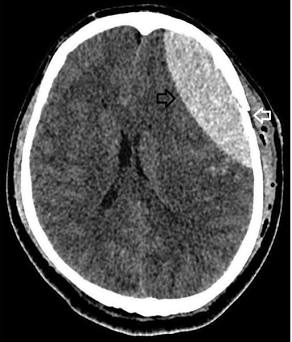
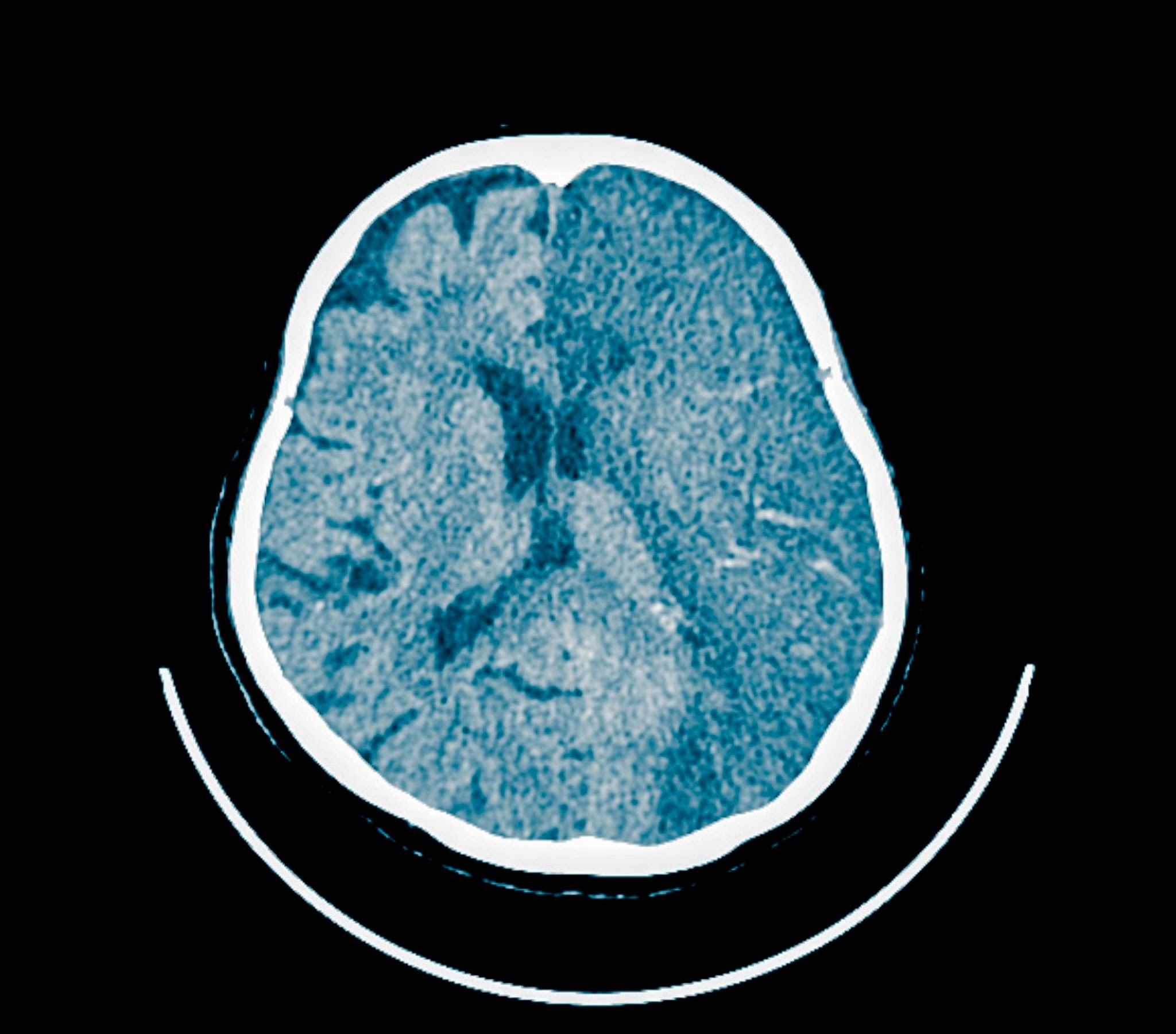
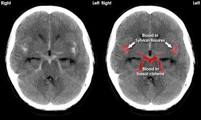
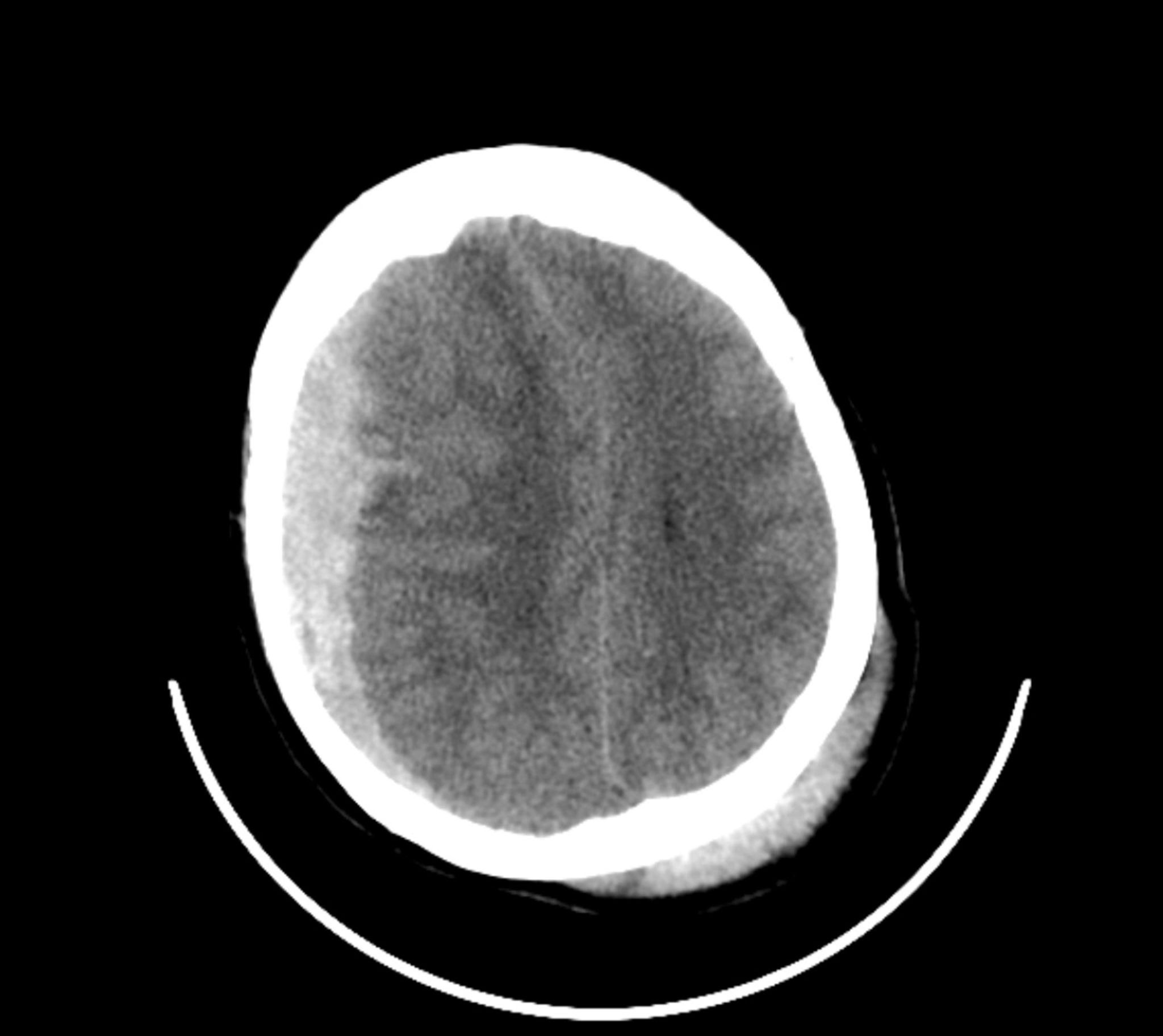
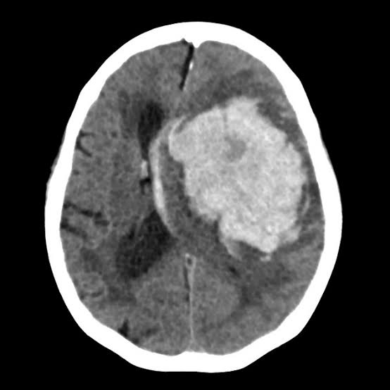
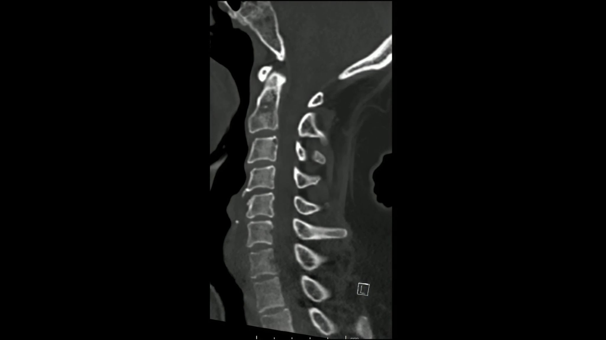
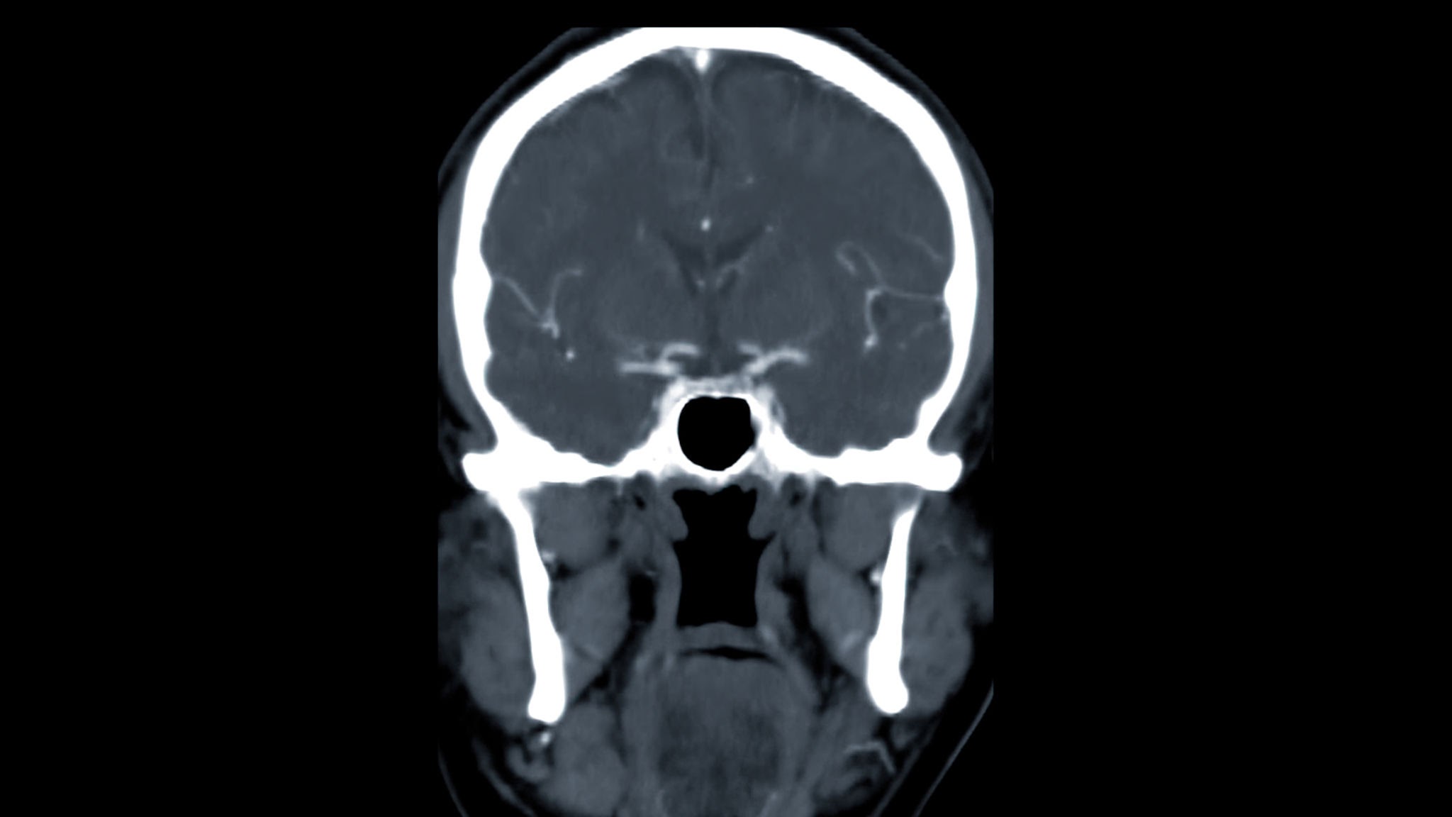
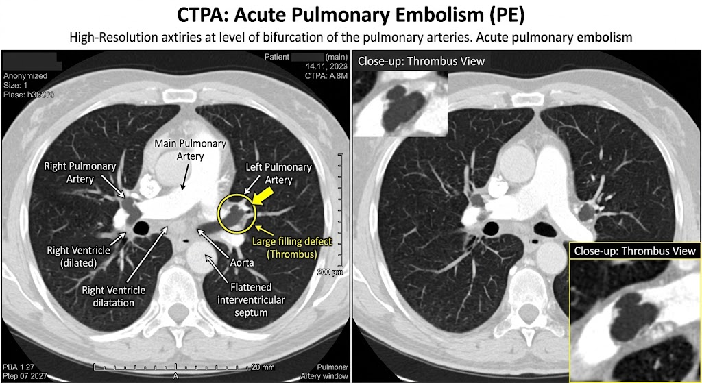
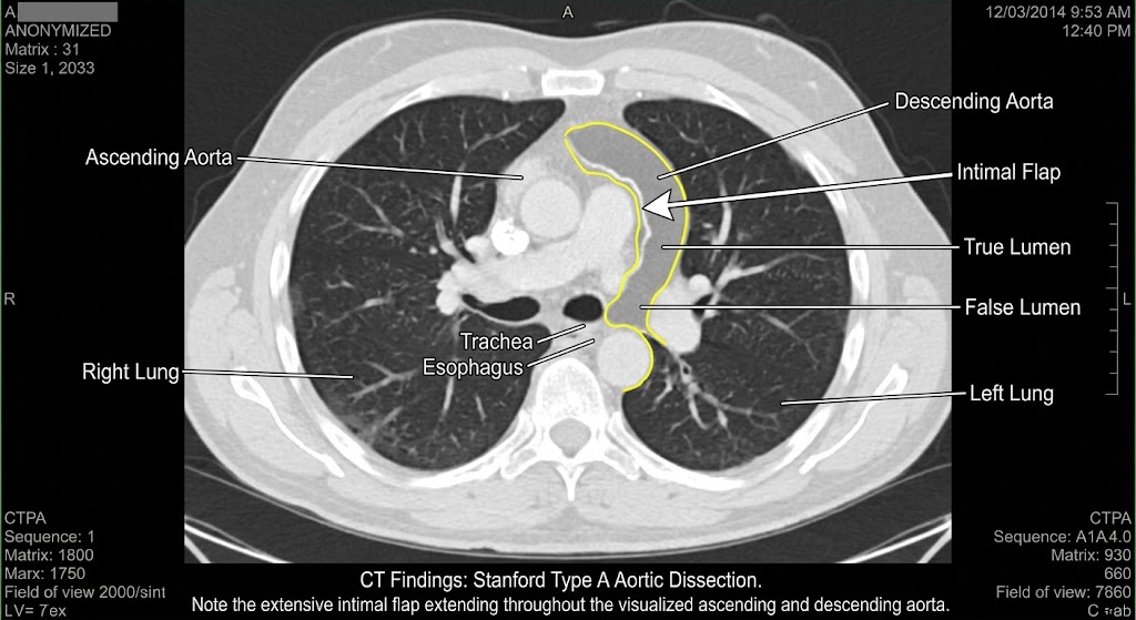
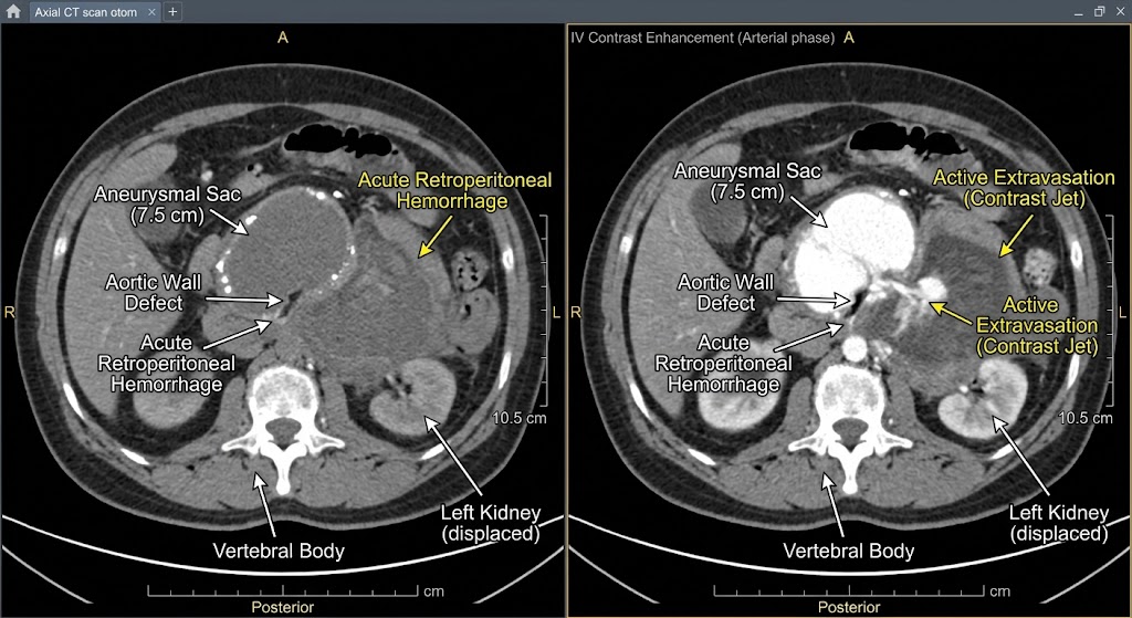
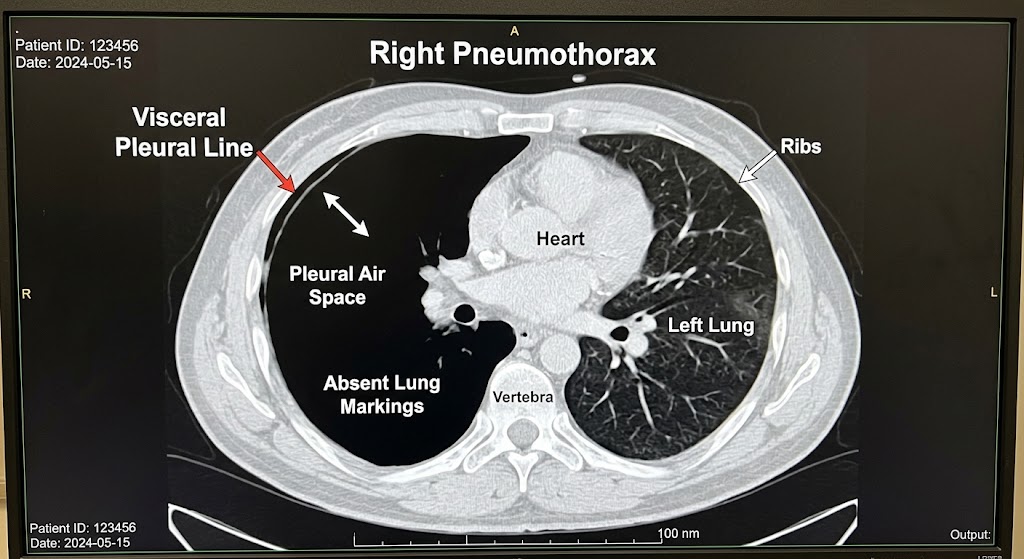
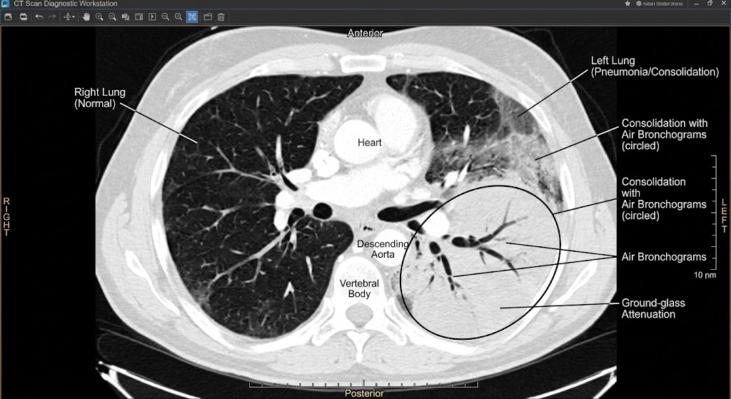

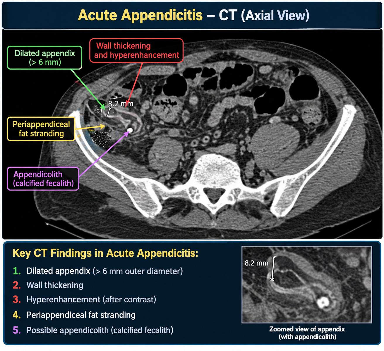
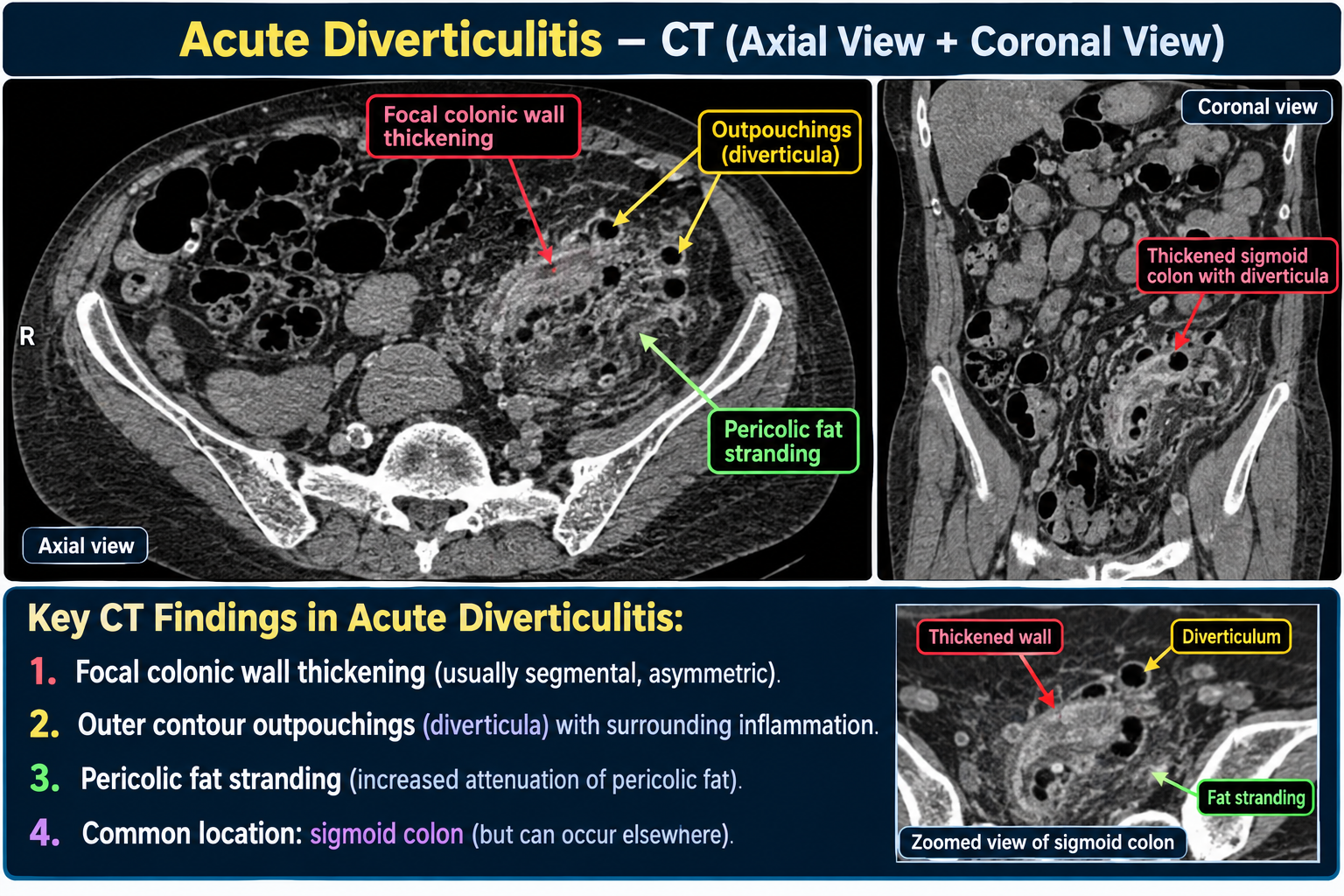
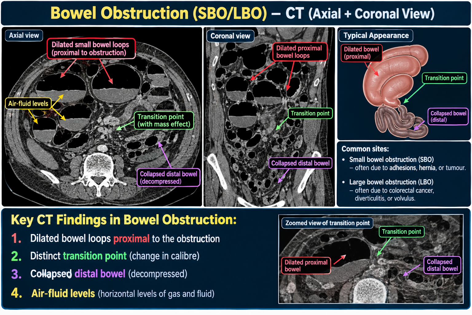
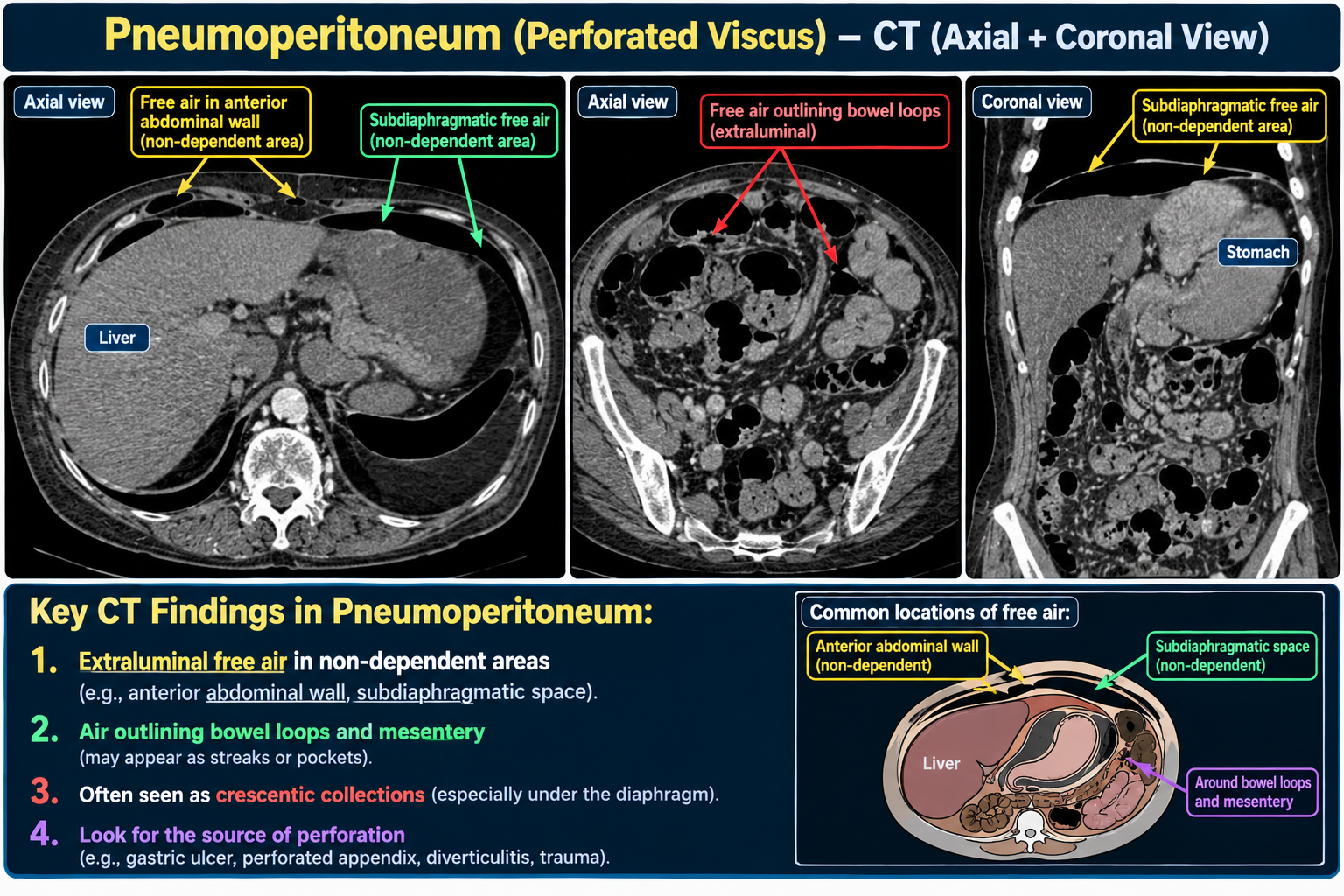
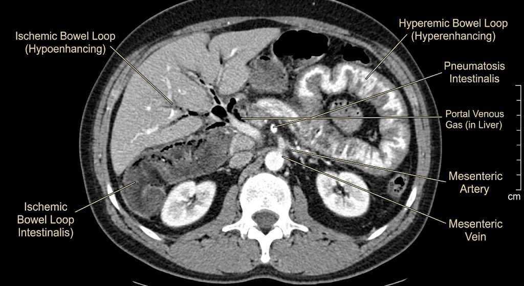
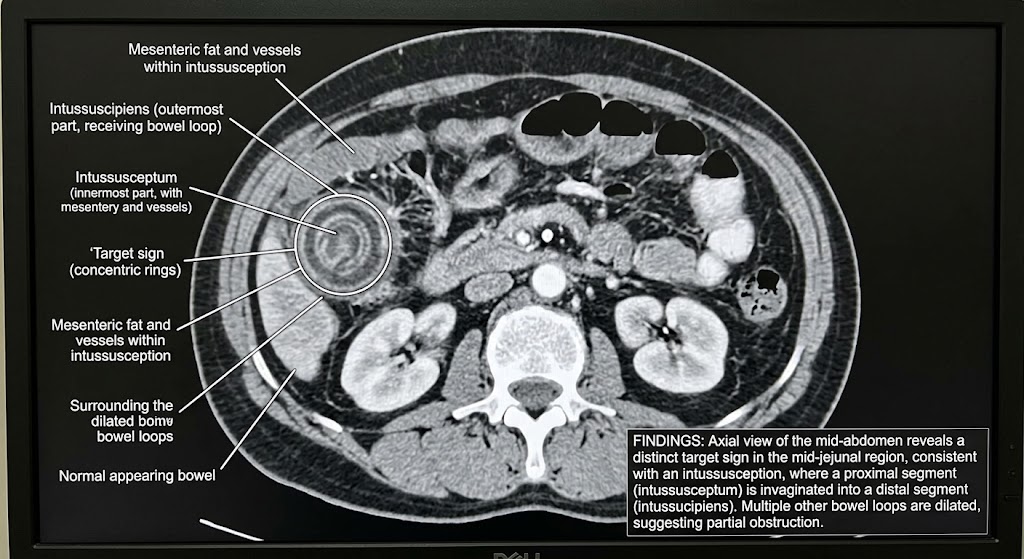
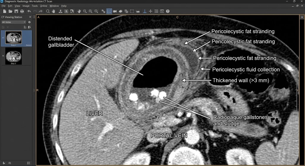
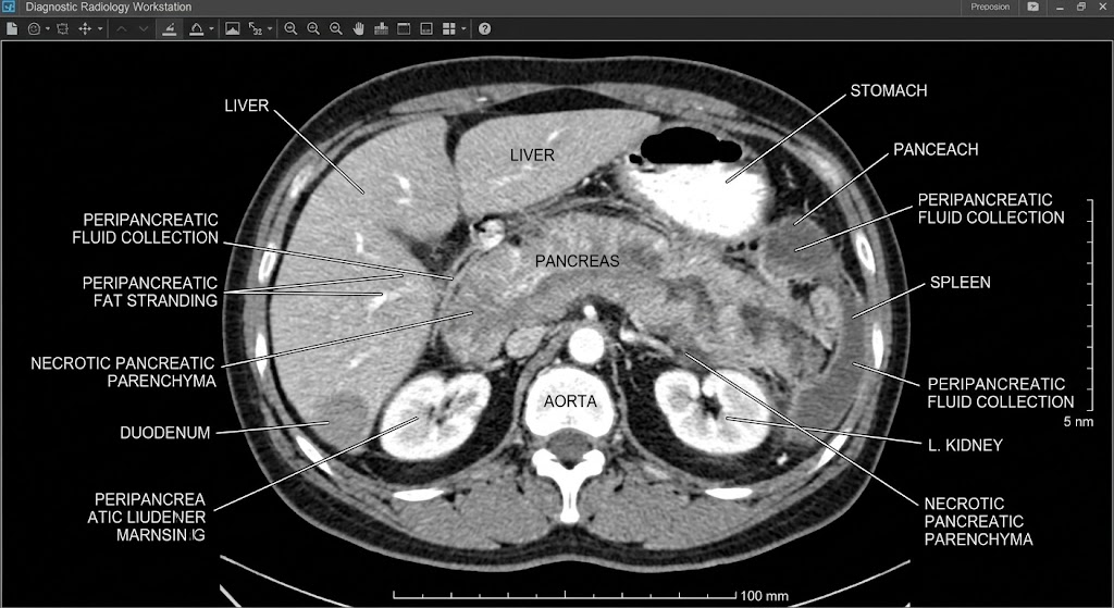 h1>Detailed Information for Acute Pancreatitis CT Scan